Copyright
©The Author(s) 2023.
World J Gastrointest Surg. Dec 27, 2023; 15(12): 2809-2819
Published online Dec 27, 2023. doi: 10.4240/wjgs.v15.i12.2809
Published online Dec 27, 2023. doi: 10.4240/wjgs.v15.i12.2809
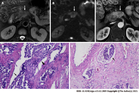
Figure 1 A 58-year-old man with pancreatic neuroendocrine tumor grade 2 underwent magnetic resonance imaging.
A-C: Magnetic resonance imaging images demonstrated the regular tumor characteristics on T2-weighted imaging (A), diffusion-weighted imaging (B) and arterial phase (C) of contrast enhanced images. The tumor shows ill-defined nodular tumor-pancreas interface with infiltrative to adjacent normal pancreatic parenchyma; D: Hematoxylin-eosin (HE) staining (× 400) reveal that the nerve (black arrowhead) is invaded by the tumor (black arrow); E: HE staining (× 400) shows that the tumor (black arrow) is located in the vascular lumen.
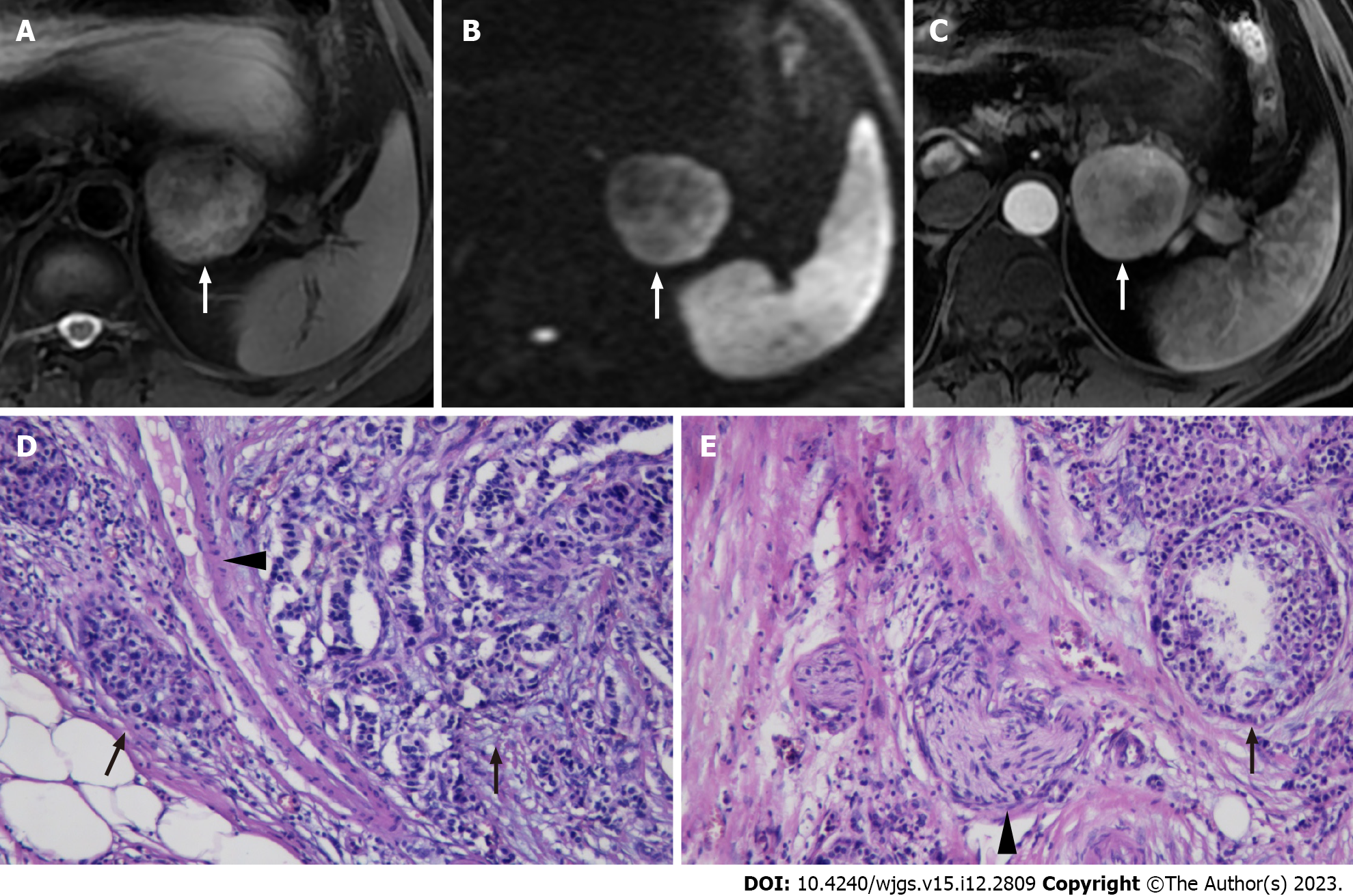
Figure 2 A 52-year-old woman with pancreatic neuroendocrine tumor grade 1 underwent magnetic resonance imaging.
A-C: Magnetic resonance imaging (MRI) images revealed the regular tumor characteristics on T2-weighted imaging (A), diffusion-weighted imaging (B) and arterial phase (C) of contrast enhanced images. A round, well demarcated tumor with smooth contours is shown on MRIs (white arrow). The tumor pancreas interface is clear; D and E: Hematoxylin-eosin staining (× 400) reveal that the space between the vascular and nerve (black arrow) and the tumor (black arrow) is clear.
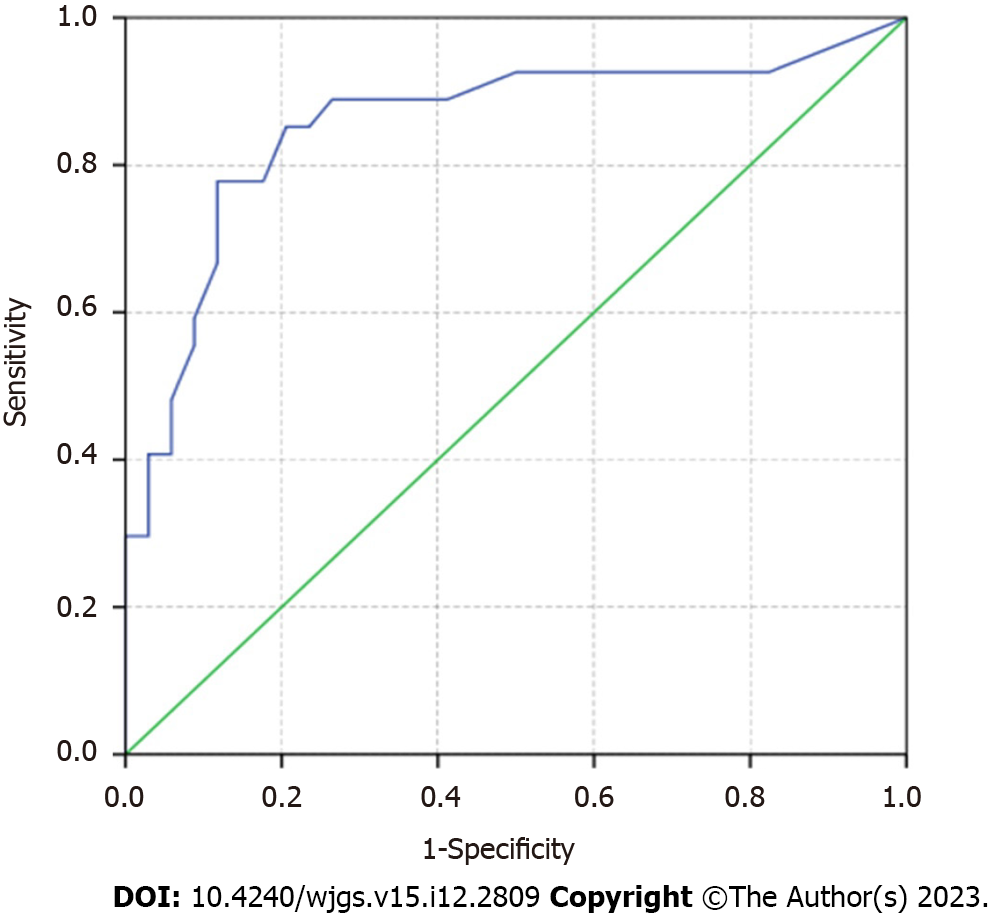
Figure 3 The receiver operating characteristic curve for diagnostic performance of multivariate model regarding the lymphatic, microvascular, and perineural invasion of pancreatic neuroendocrine tumors.
The area under the curve is 0.855 (95% confidence interval: 0.750-0.960).
- Citation: Liu YL, Zhu HB, Chen ML, Sun W, Li XT, Sun YS. Prediction of the lymphatic, microvascular, and perineural invasion of pancreatic neuroendocrine tumors using preoperative magnetic resonance imaging. World J Gastrointest Surg 2023; 15(12): 2809-2819
- URL: https://www.wjgnet.com/1948-9366/full/v15/i12/2809.htm
- DOI: https://dx.doi.org/10.4240/wjgs.v15.i12.2809


