Copyright
©The Author(s) 2023.
World J Gastrointest Surg. Dec 27, 2023; 15(12): 2693-2708
Published online Dec 27, 2023. doi: 10.4240/wjgs.v15.i12.2693
Published online Dec 27, 2023. doi: 10.4240/wjgs.v15.i12.2693
Figure 1 Annual publication trend of articles related to indocyanine green fluorescence.
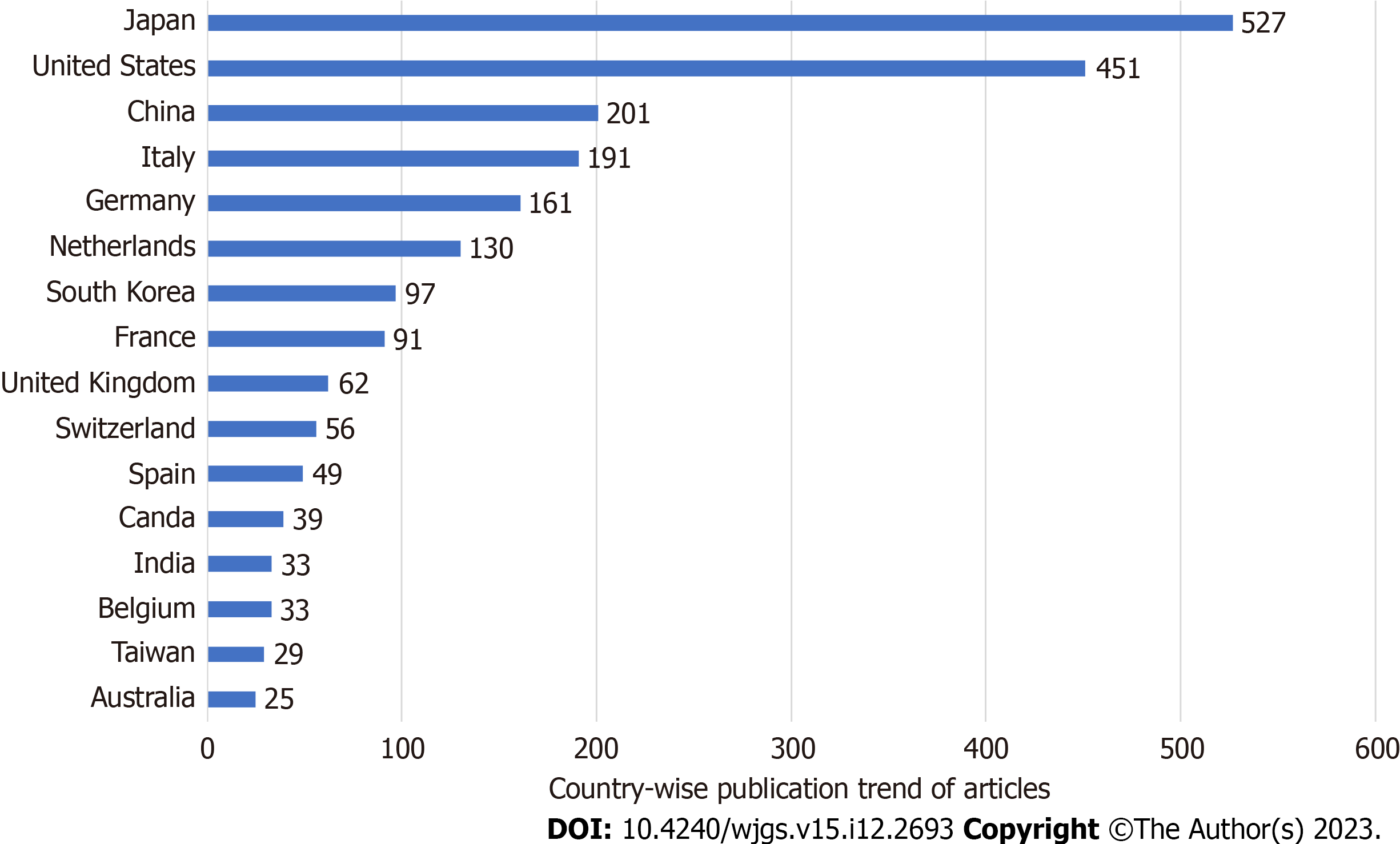
Figure 2 Country-wise publication trend of articles related to indocyanine green fluorescence.
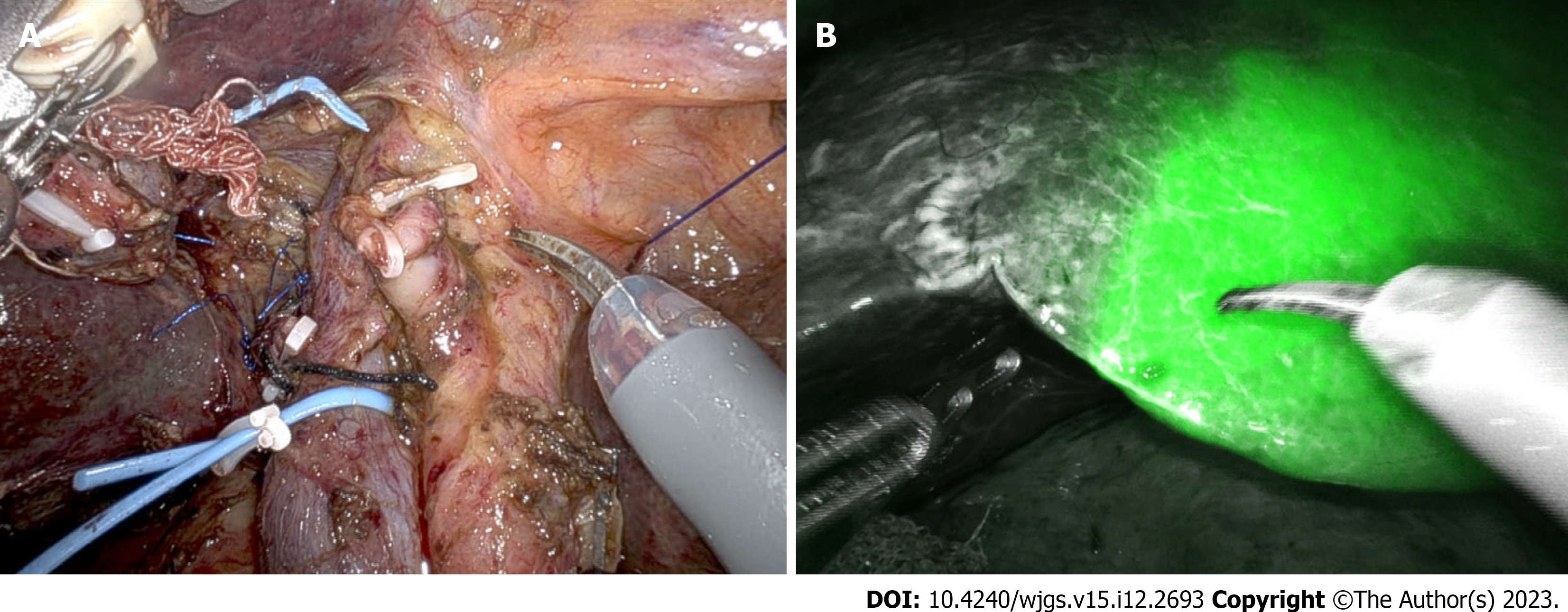
Figure 3 Negative indocyanine green staining technique used in a patient with hilar cholangiocarcinoma planned for right hepatectomy with caudate lobectomy.
A: Portal and hepatic artery branches of the right hemiliver to be resected are ligated; B: Line of transection for right hepatectomy identified with the help of indocyanine green fluorescence.
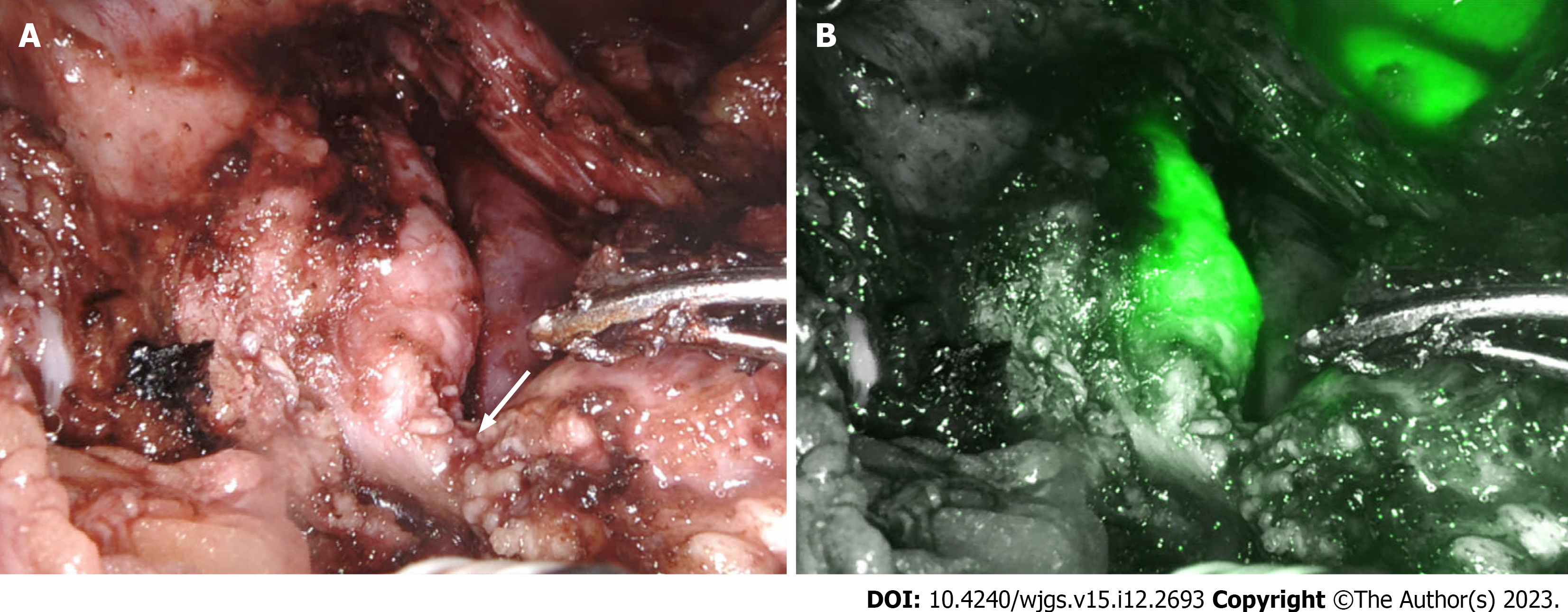
Figure 4 Identification of bile duct during robotic duodenum preserving pancreas head resection.
A: Pancreatic duct (arrow) divided at its junction with the bile duct; B: Indocyanine green fluorescence demonstrates bile duct.
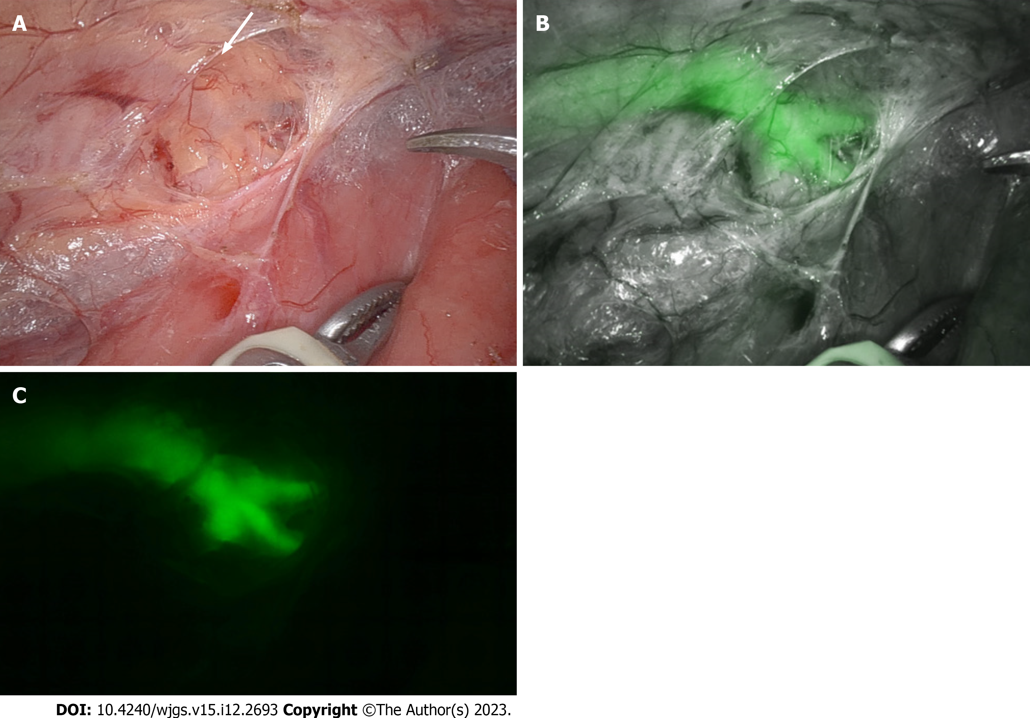
Figure 5 Visualization of the thoracic duct.
A and B: Visualization of the thoracic duct during robotic esophagectomy in normal mode (A) is enhanced by indocyanine green fluorescence (B); C: The branching pattern of the thoracic duct is well visualized in fluorescence mode.
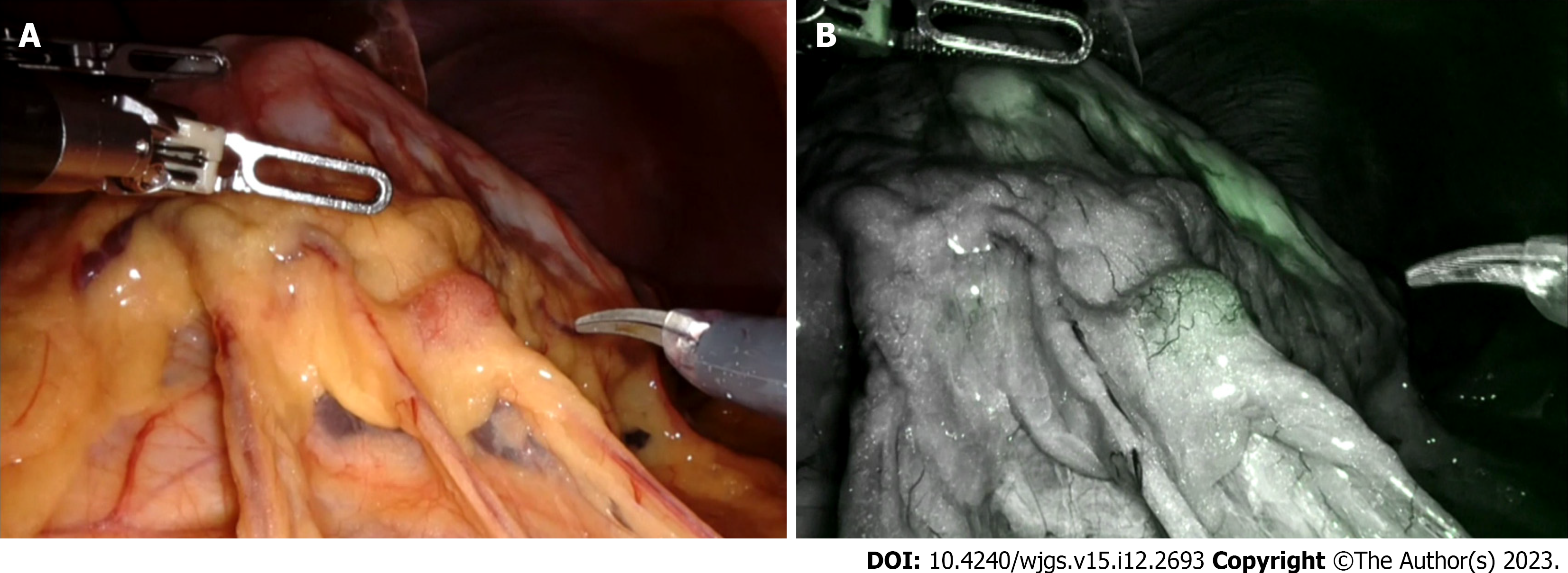
Figure 6 Visualization of perigastric lymph node during robotic D2 gastrectomy in normal mode and fluorescence mode.
A: In normal mode; B: In fluorescence mode.
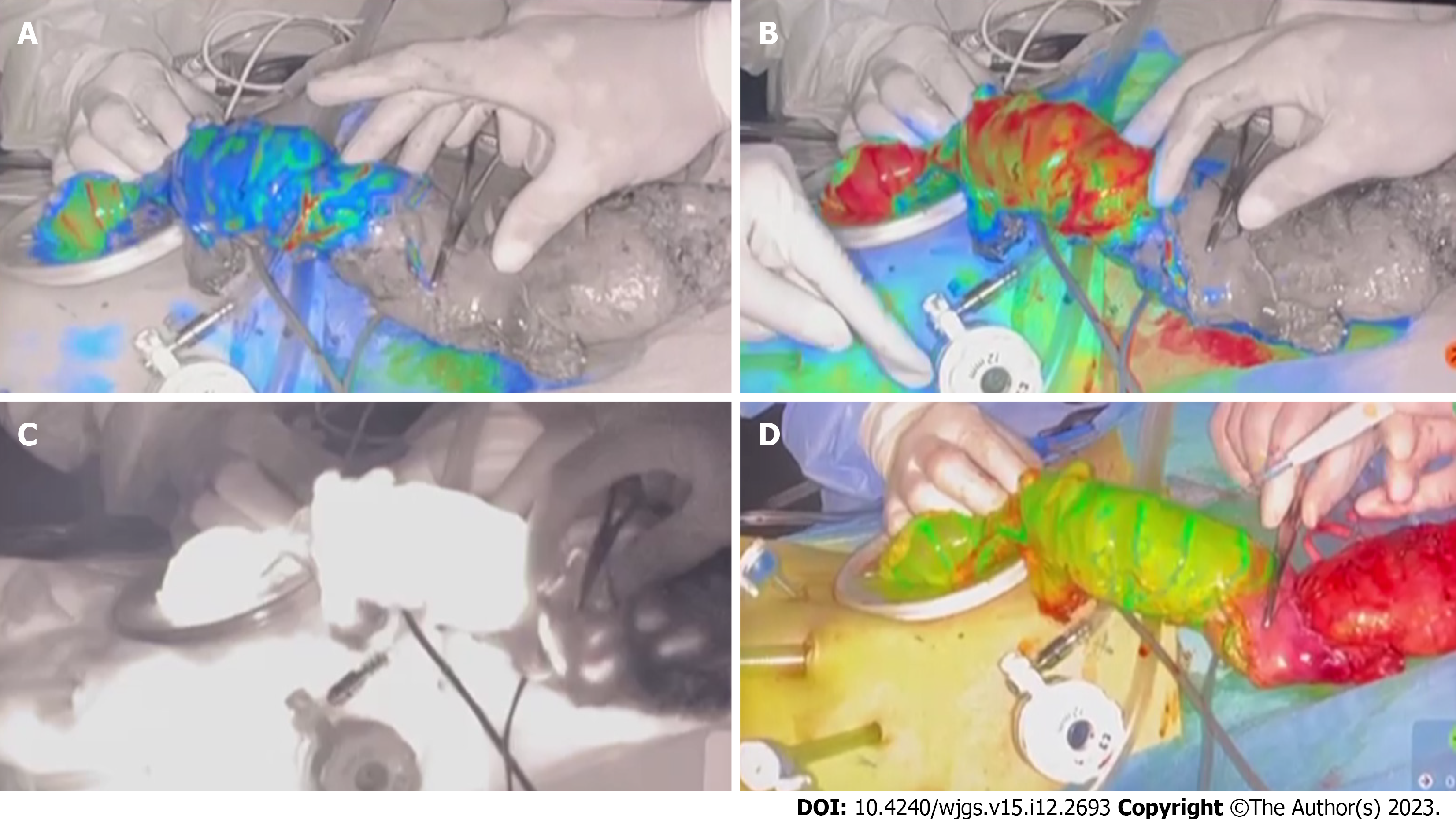
Figure 7 Objective evaluation of colonic perfusion using indocyanine green fluorescence during low anterior resection.
A and B: Increasing fluorescence levels show the transition from blue (A) to red (B) and guide the selection of the line of bowel division; C: Fluorescence is displayed on a Grayscale; D: Fluorescence displayed in overlay mode.
- Citation: Kalayarasan R, Chandrasekar M, Sai Krishna P, Shanmugam D. Indocyanine green fluorescence in gastrointestinal surgery: Appraisal of current evidence. World J Gastrointest Surg 2023; 15(12): 2693-2708
- URL: https://www.wjgnet.com/1948-9366/full/v15/i12/2693.htm
- DOI: https://dx.doi.org/10.4240/wjgs.v15.i12.2693


