Copyright
©The Author(s) 2023.
World J Gastrointest Surg. Oct 27, 2023; 15(10): 2367-2375
Published online Oct 27, 2023. doi: 10.4240/wjgs.v15.i10.2367
Published online Oct 27, 2023. doi: 10.4240/wjgs.v15.i10.2367
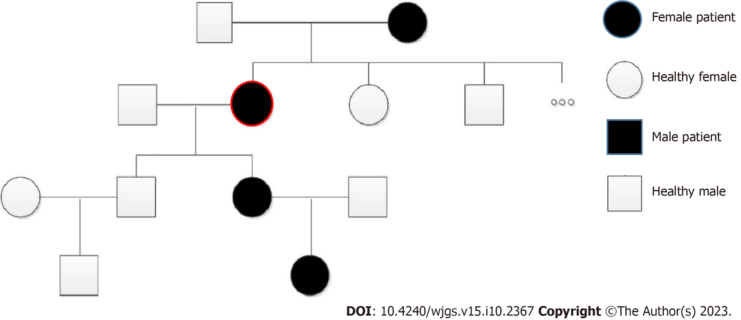
Figure 1 Pedigree chart.
Black circles refer to female patients with a history of epistaxis, open circles refer to healthy women; black squares refer to male patients with a history of epistaxis, open squares refer to healthy men. Red circle highlights the patient in this case study.
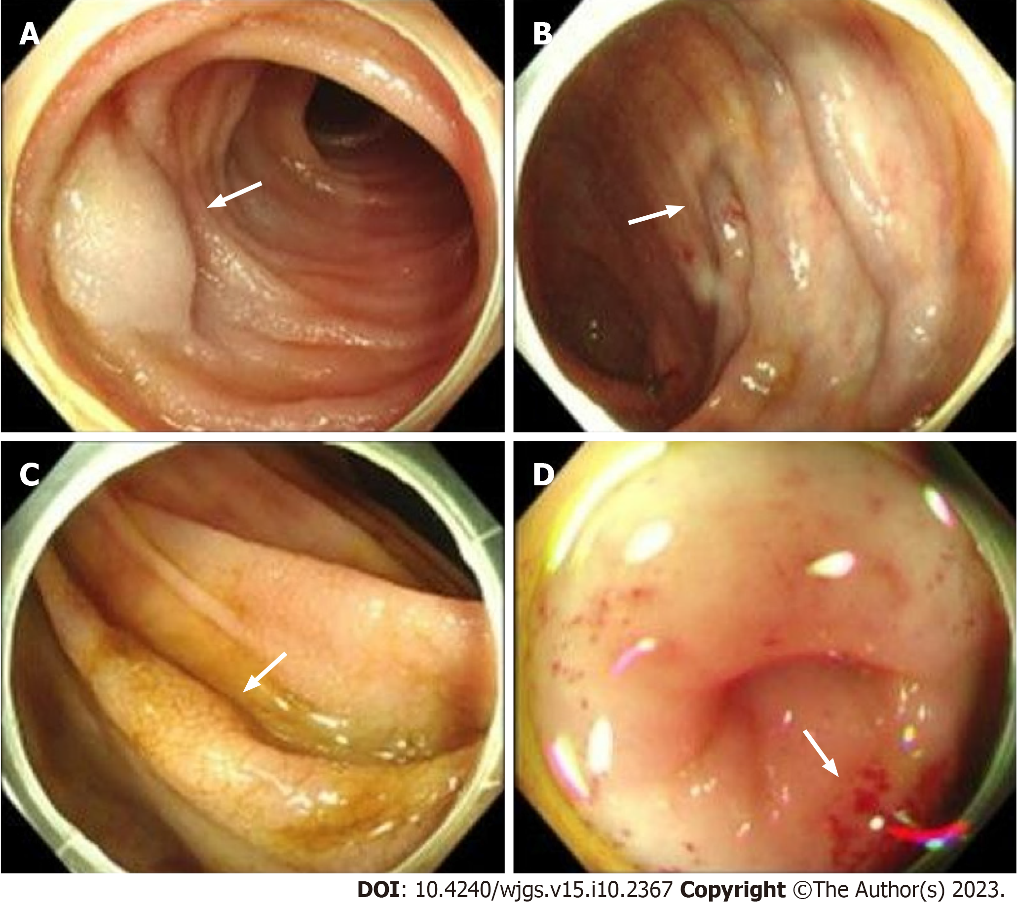
Figure 2 Endoscopy images.
A: Aneurysm (arrows) on intestinal mucosa; B: Varicose vein (arrows) on intestinal mucosa; C: Cracked and scale-like mucosa (arrows) on intestinal mucosa; D: Scattered sites of bleeding sites (arrows) on intestinal mucosa.
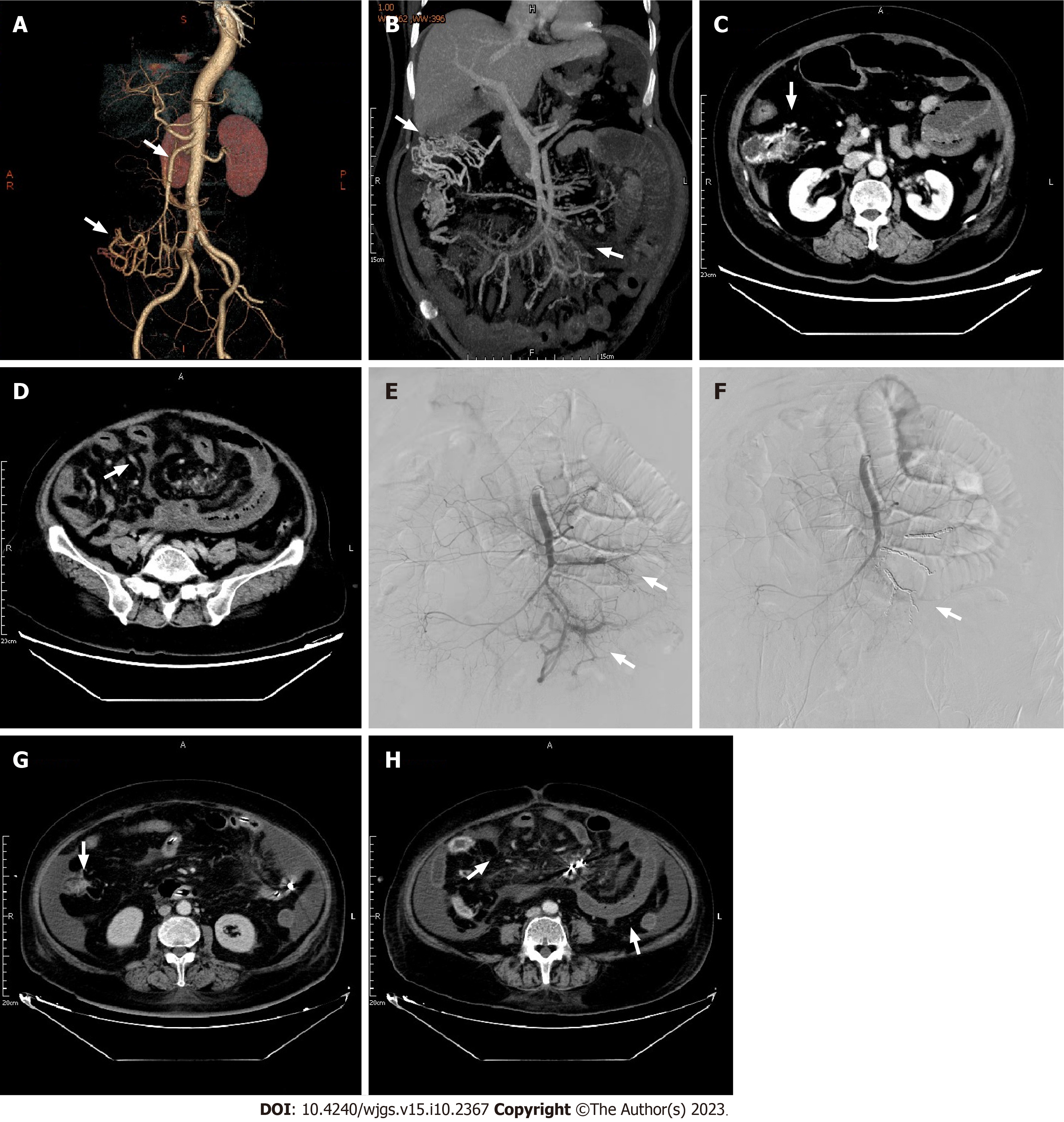
Figure 3 Pre- and post-procedure computed tomography images.
A: Computed tomography (CT) angiography examination of abdominal blood vessels. The superior mesenteric vein (upper arrow), and tortuous blood vessels connecting with the superior mesenteric vein (lower arrow); B: Tortuous vessels (left arrow), and the superior mesenteric arteriovenous fistula (right arrow); C: CT examination. Tortuous vessels (arrow); D: Considerable edema of the intestinal segment receiving blood supply from the jejunal and ileal branches of the superior mesenteric artery (arrow); E: Two arteriovenous fistulas (arrows); F-H: After embolization, arteriovenous fistulas were not present (F), the range of tortuous vessels was decreased (G), and edema of the intestinal segment receiving blood supply from the jejunal and ileal branches of the superior mesenteric artery was relieved (H).
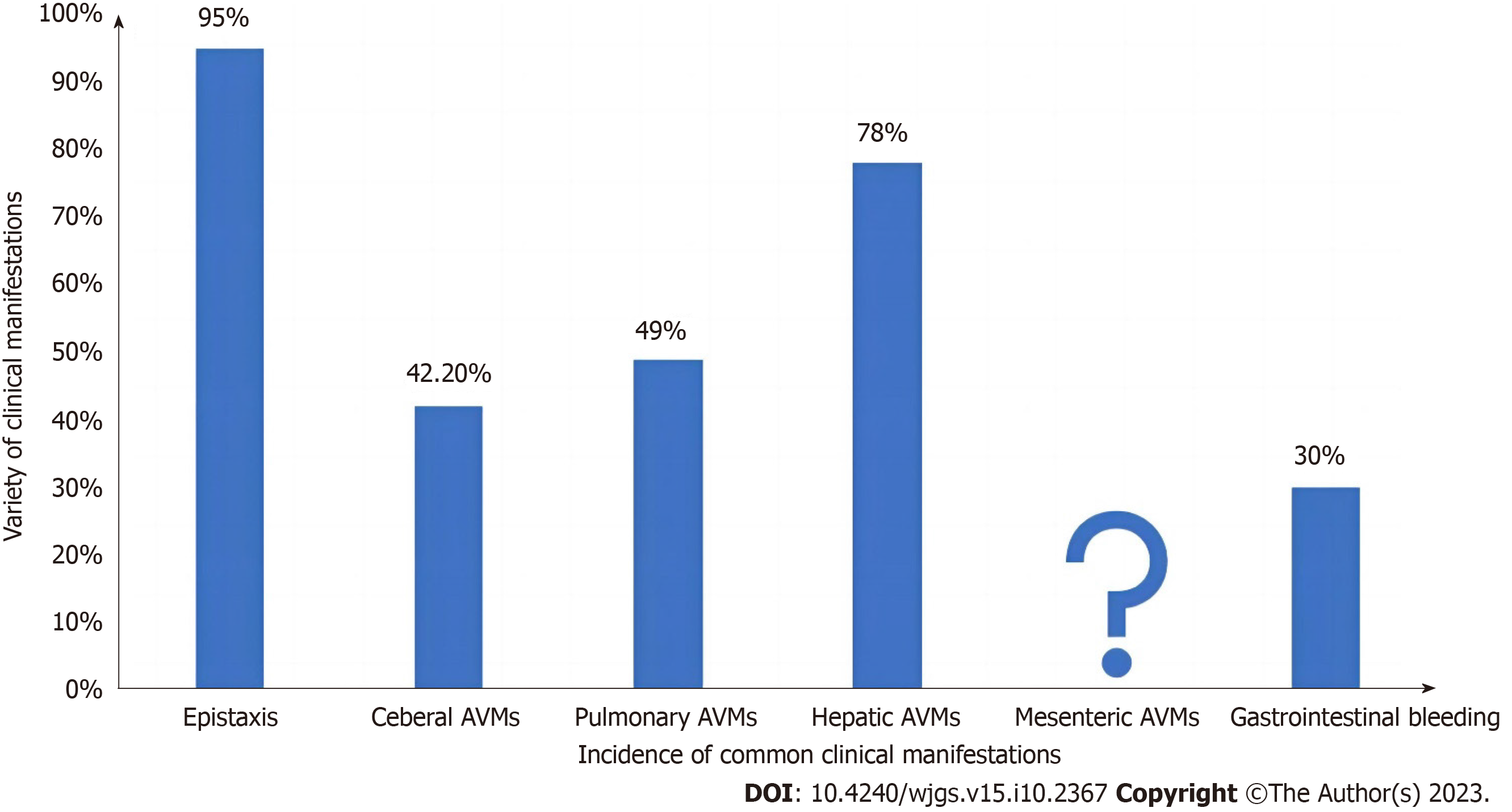
Figure 4 Common clinical manifestations of hereditary hemorrhagic telangiectasia patients.
AVM: Arteriovenous malformation; HHT: Hereditary hemorrhagic telangiectasia.
- Citation: Wu JL, Zhao ZZ, Chen J, Zhang HW, Luan Z, Li CY, Zhao YM, Jing YJ, Wang SF, Sun G. Hereditary hemorrhagic telangiectasia involving portal venous system: A case report and review of the literature. World J Gastrointest Surg 2023; 15(10): 2367-2375
- URL: https://www.wjgnet.com/1948-9366/full/v15/i10/2367.htm
- DOI: https://dx.doi.org/10.4240/wjgs.v15.i10.2367


