Copyright
©The Author(s) 2023.
World J Gastrointest Surg. Oct 27, 2023; 15(10): 2098-2107
Published online Oct 27, 2023. doi: 10.4240/wjgs.v15.i10.2098
Published online Oct 27, 2023. doi: 10.4240/wjgs.v15.i10.2098
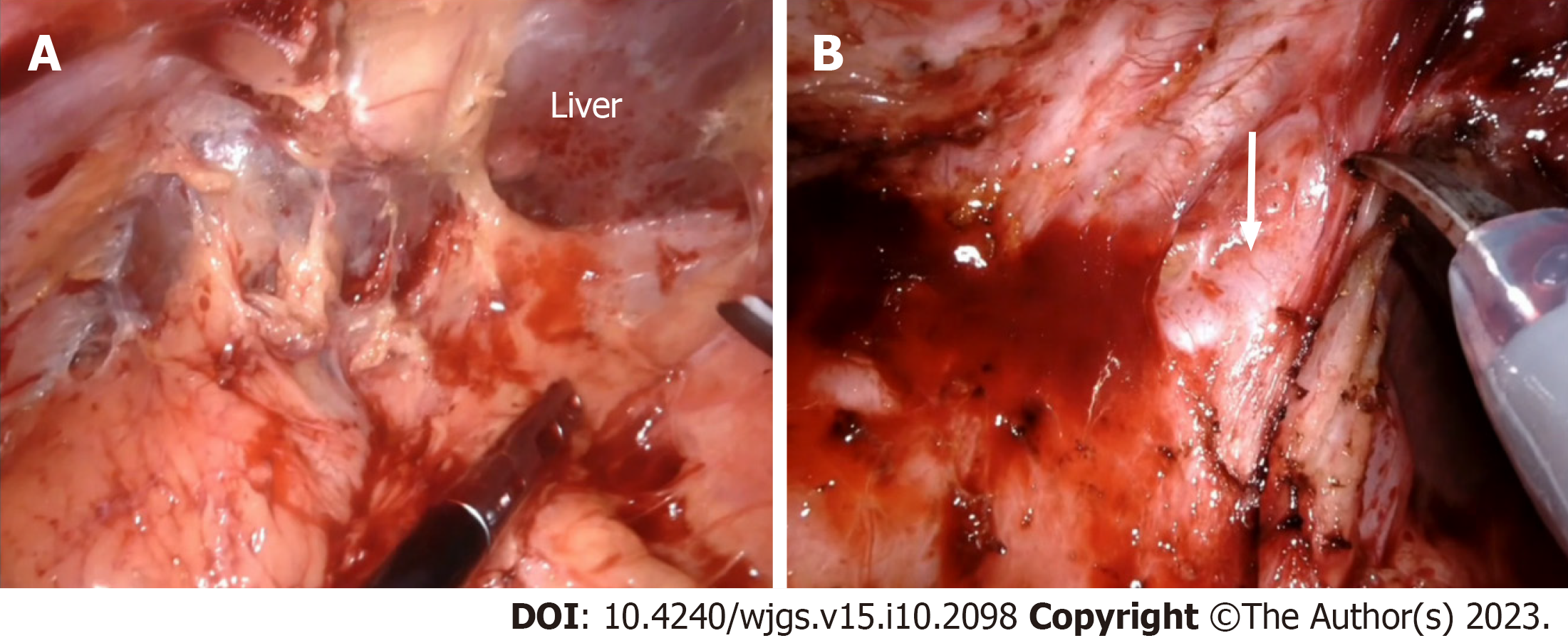
Figure 1 Adhesiolysis and initial dissection phase.
A: Perihepatic adhesions are left undisturbed to facilitate liver retraction and exposure of the hilum; B: Dissection proceeds towards the umbilical fissure with careful identification and preservation of the left hepatic artery (arrow).
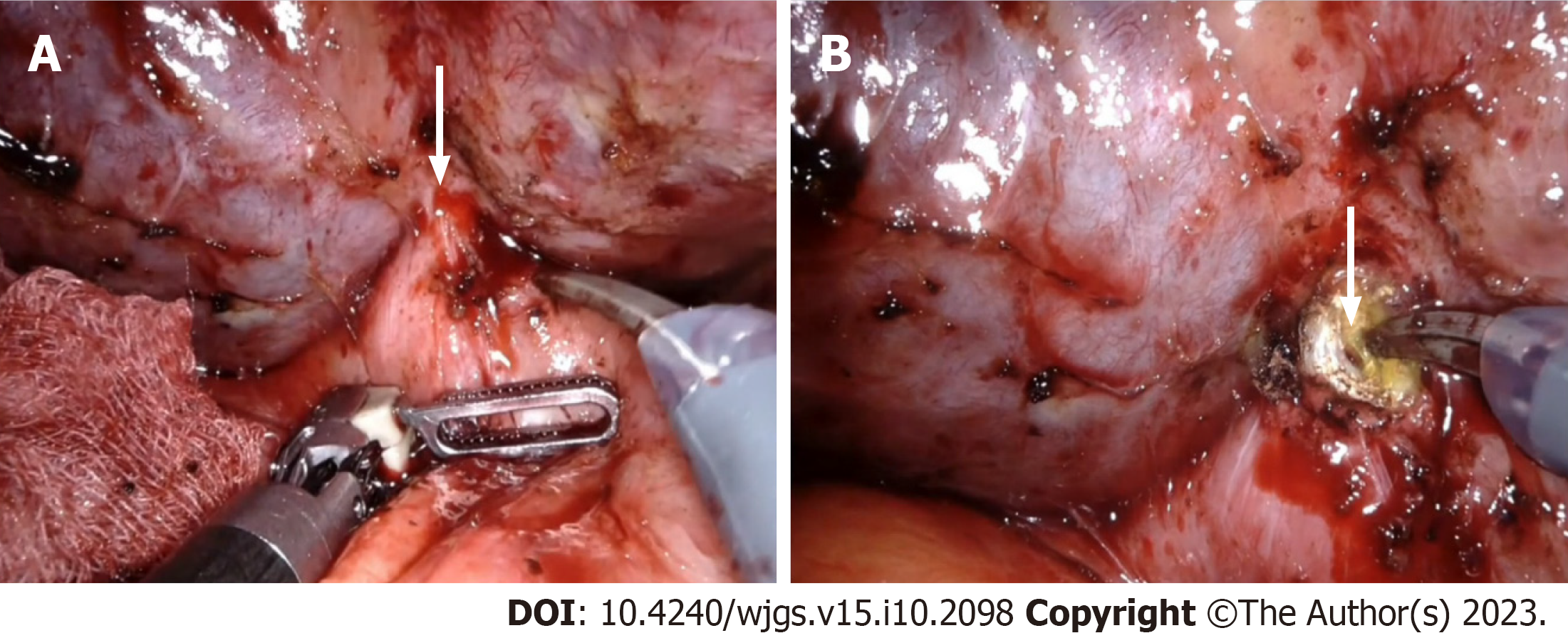
Figure 2 Identification of hepatic duct.
A: Internal fistula between the hepatic duct and duodenum (arrow); B: Division of the fistula facilitates visualization of the hepatic duct (arrow).
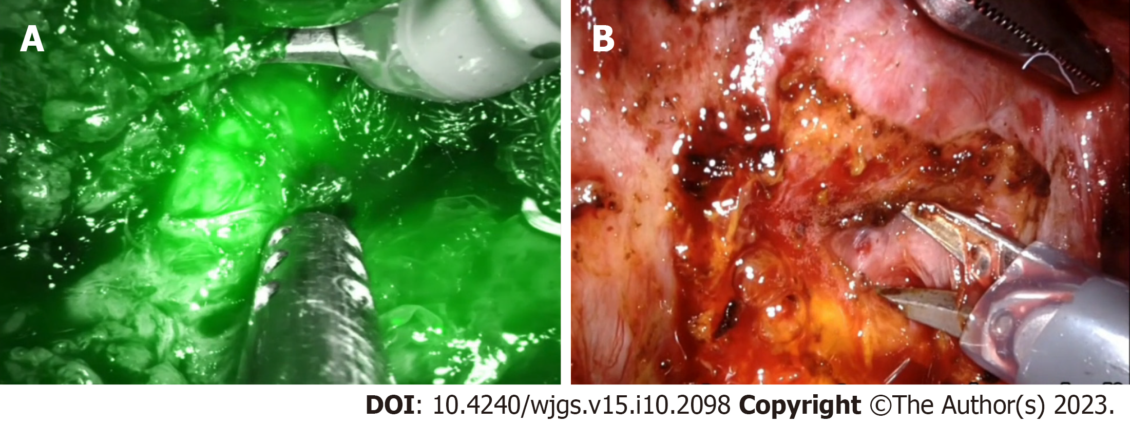
Figure 3 Lowering the hilar plate.
A: Indocyanine green fluorescence facilitates hepatic duct identification; B: Hilar plate lowered by dissection between the Glissonean sheath and Laennec’s capsule.
Figure 4 Opening the hepatic duct.
A: Identification and opening of the left hepatic duct; B: Confluence of left hepatic duct with right hepatic duct identified.
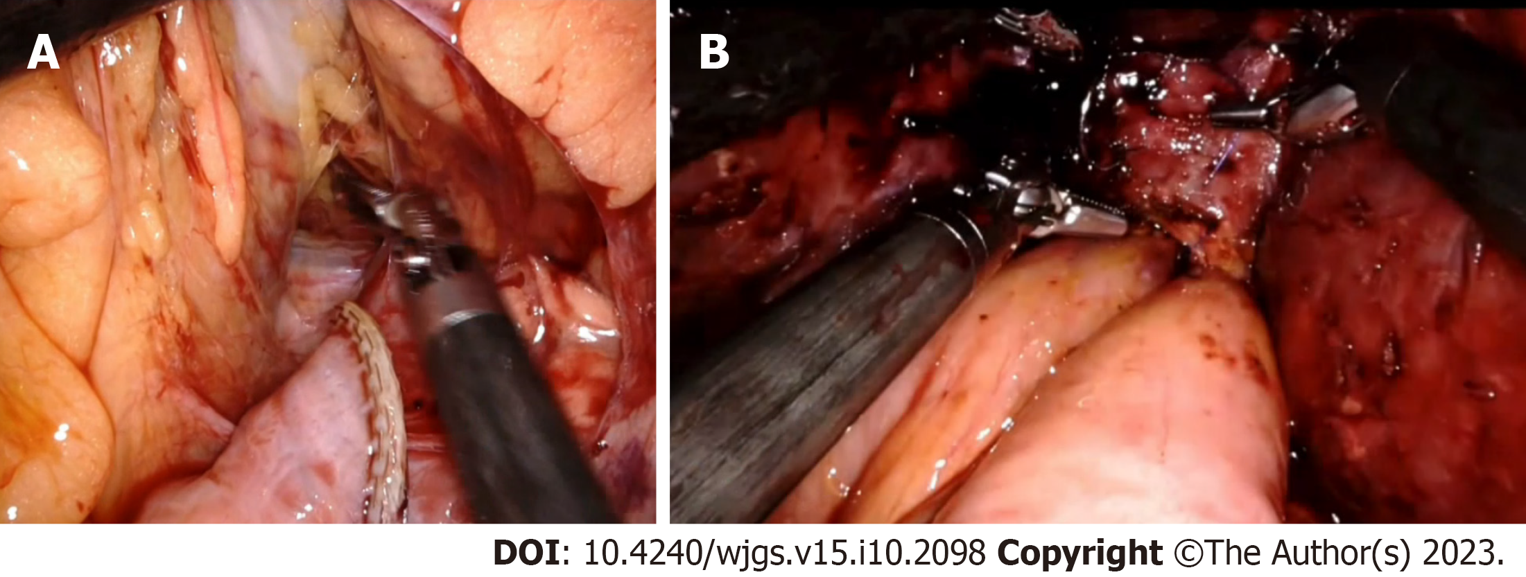
Figure 5 Roux-en-Y hepaticojejunostomy.
A: Roux limb of jejunum taken to the supracolic compartment through the mesocolic window; B: Completed hepaticojejunostomy.
- Citation: Kalayarasan R, Sai Krishna P. Minimally invasive surgery for post cholecystectomy biliary stricture: current evidence and future perspectives. World J Gastrointest Surg 2023; 15(10): 2098-2107
- URL: https://www.wjgnet.com/1948-9366/full/v15/i10/2098.htm
- DOI: https://dx.doi.org/10.4240/wjgs.v15.i10.2098


