Copyright
©The Author(s) 2022.
World J Gastrointest Surg. May 27, 2022; 14(5): 374-382
Published online May 27, 2022. doi: 10.4240/wjgs.v14.i5.374
Published online May 27, 2022. doi: 10.4240/wjgs.v14.i5.374
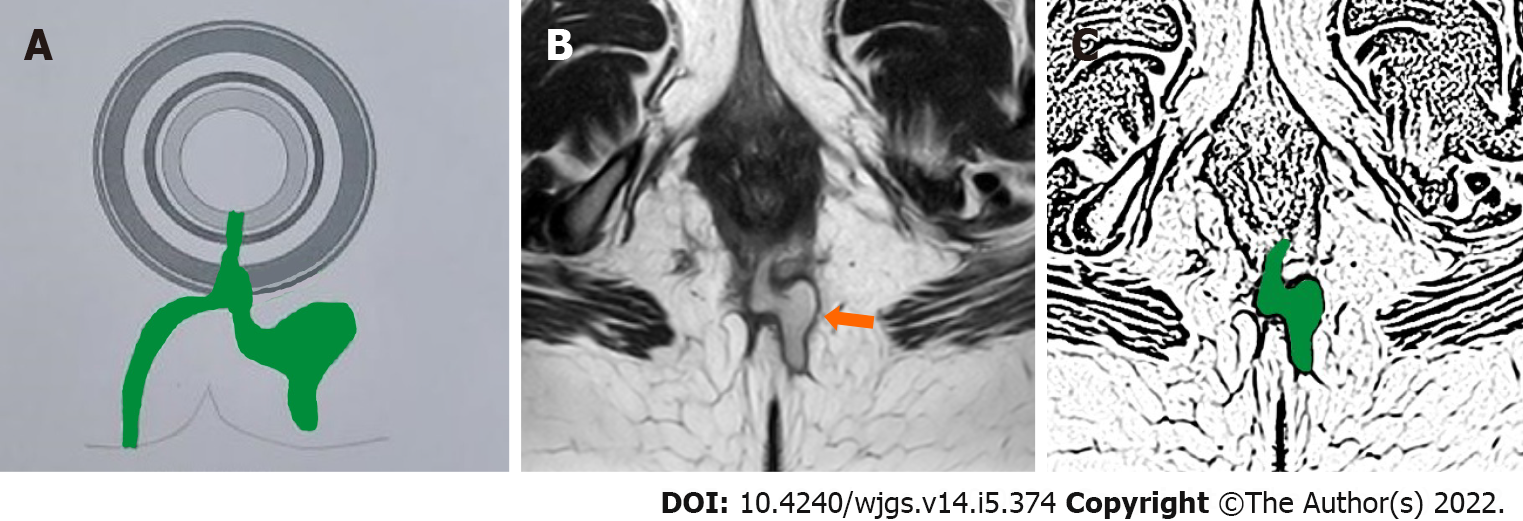
Figure 1 A 43-year-old female patient with recurrent high transsphincteric posterior anal fistula with multiple branches.
The intersphincteric component of fistula is a single linear tract at 6 o’clock (posterior) and the rest of all the fistula tracts are outside the external sphincter. This fistula is better managed by ligation of intersphincteric fistula tract procedure. A: Axial section-schematic diagram; B: T2-weighted magnetic resonance imaging axial section (orange arrow pointing the fistula tract); C: Sketch of B (fistula tract being shown in green color).
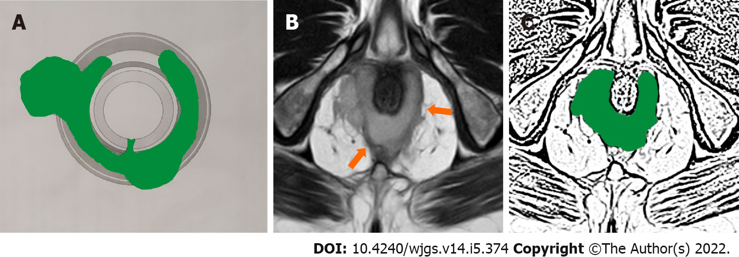
Figure 2 A 47-year-old male patient with high posterior intersphincteric anal fistula with abscess.
This fistula is difficult to manage by ligation of intersphincteric fistula tract and is better managed by transanal opening of intersphincteric space procedure. A: Axial section-schematic diagram; B: T2-weighted magnetic resonance imaging axial section (orange arrows pointing the fistula tract); C: Sketch of B (fistula tract being shown in green color).
Figure 3 A 39-year-old male patient with right sided suprasphincteric anal fistula with abscess.
This fistula is difficult to manage by ligation of intersphincteric fistula tract and is better managed by transanal opening of intersphincteric space procedure. A: Coronal section-schematic diagram; B: T2-weighted magnetic resonance imaging coronal section (orange arrows pointing the fistula tract); C: Sketch of B (fistula tract being shown in green color).
- Citation: Garg P. Comparison between recent sphincter-sparing procedures for complex anal fistulas-ligation of intersphincteric tract vs transanal opening of intersphincteric space. World J Gastrointest Surg 2022; 14(5): 374-382
- URL: https://www.wjgnet.com/1948-9366/full/v14/i5/374.htm
- DOI: https://dx.doi.org/10.4240/wjgs.v14.i5.374


