Copyright
©The Author(s) 2017.
World J Diabetes. Apr 15, 2017; 8(4): 135-142
Published online Apr 15, 2017. doi: 10.4239/wjd.v8.i4.135
Published online Apr 15, 2017. doi: 10.4239/wjd.v8.i4.135
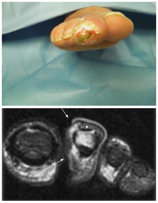
Figure 5 Osteomyelitis of second toe (distal phalanx) revealed by magnetic resonance imaging.
The arrows and the arrowhead show the bone involvement of distal phalanx (second toe).
- Citation: Giurato L, Meloni M, Izzo V, Uccioli L. Osteomyelitis in diabetic foot: A comprehensive overview. World J Diabetes 2017; 8(4): 135-142
- URL: https://www.wjgnet.com/1948-9358/full/v8/i4/135.htm
- DOI: https://dx.doi.org/10.4239/wjd.v8.i4.135


