Copyright
©The Author(s) 2016.
World J Diabetes. Jul 10, 2016; 7(13): 271-278
Published online Jul 10, 2016. doi: 10.4239/wjd.v7.i13.271
Published online Jul 10, 2016. doi: 10.4239/wjd.v7.i13.271
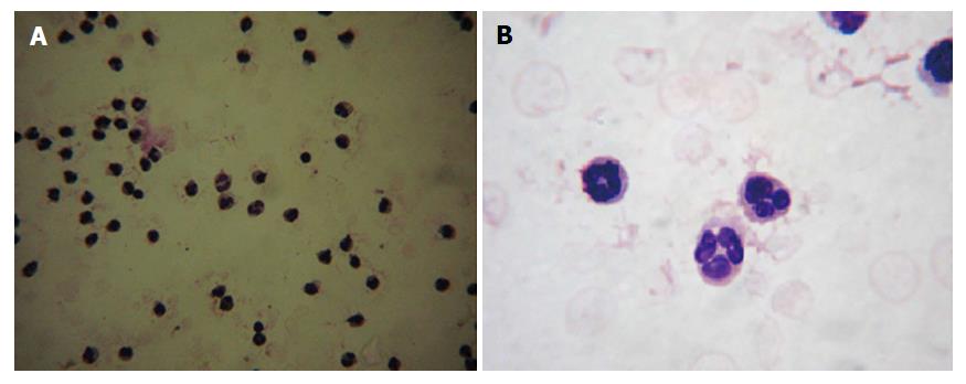
Figure 1 Morphological analysis of isolated neutrophils.
Isolated neutrophils were smeared on a glass slide and labeled with Leishman stain. Under the 20 × magnification, cells appeared homogeneous (A) and 40 × magnification exhibited the multi-lobulated nucleus (B).
- Citation: Ridzuan N, John CM, Sandrasaigaran P, Maqbool M, Liew LC, Lim J, Ramasamy R. Preliminary study on overproduction of reactive oxygen species by neutrophils in diabetes mellitus. World J Diabetes 2016; 7(13): 271-278
- URL: https://www.wjgnet.com/1948-9358/full/v7/i13/271.htm
- DOI: https://dx.doi.org/10.4239/wjd.v7.i13.271


