Copyright
©2014 Baishideng Publishing Group Inc.
World J Diabetes. Dec 15, 2014; 5(6): 882-888
Published online Dec 15, 2014. doi: 10.4239/wjd.v5.i6.882
Published online Dec 15, 2014. doi: 10.4239/wjd.v5.i6.882
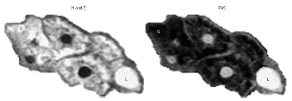
Figure 2 Schematic reproduction of staining with Hematoxilyn and Eosin vs Periodic Acid Schiff.
The glycogen (G) disappears in H and E whereas it stains (red) in PAS. The presence of lipids (L) in focal vescicular steatosis is demonstrated by lack of staining both in HE and PAS. H and E: Hematoxilyn and Eosin; PAS: Periodic Acid Schiff.
- Citation: Giordano S, Martocchia A, Toussan L, Stefanelli M, Pastore F, Devito A, Risicato MG, Ruco L, Falaschi P. Diagnosis of hepatic glycogenosis in poorly controlled type 1 diabetes mellitus. World J Diabetes 2014; 5(6): 882-888
- URL: https://www.wjgnet.com/1948-9358/full/v5/i6/882.htm
- DOI: https://dx.doi.org/10.4239/wjd.v5.i6.882


