Copyright
©2014 Baishideng Publishing Group Inc.
World J Diabetes. Oct 15, 2014; 5(5): 724-729
Published online Oct 15, 2014. doi: 10.4239/wjd.v5.i5.724
Published online Oct 15, 2014. doi: 10.4239/wjd.v5.i5.724
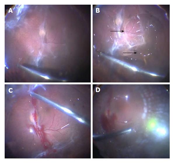
Figure 2 En bloc perfluorodissection performed in a case of tractional retinal detachment in proliferative diabetic retinopathy.
A: An opening is made with the vitrector in the mid-periphery of the posterior hyaloid; B: Perfluorocarbon liquid (PFCL) is injected to separate the posterior hyaloid from the retina (arrows). A dual bore cannula (for 23-gauge cases) attached to a 5 cc syringe filled with PFCL is used to separate membranes and posterior hyaloid from the underlying retina; C: Once all the tissues have been separated from the retina, vitrectomy can be continued up to the periphery; D: Endolaser is applied under PFCL (shown). An air-fluid and an air-gas (C3F8) exchange are performed to end the case (not shown).
- Citation: Arevalo JF, Serrano MA, Arias JD. Perfluorocarbon in vitreoretinal surgery and preoperative bevacizumab in diabetic tractional retinal detachment. World J Diabetes 2014; 5(5): 724-729
- URL: https://www.wjgnet.com/1948-9358/full/v5/i5/724.htm
- DOI: https://dx.doi.org/10.4239/wjd.v5.i5.724


