Copyright
©2014 Baishideng Publishing Group Co.
World J Diabetes. Feb 15, 2014; 5(1): 59-68
Published online Feb 15, 2014. doi: 10.4239/wjd.v5.i1.59
Published online Feb 15, 2014. doi: 10.4239/wjd.v5.i1.59
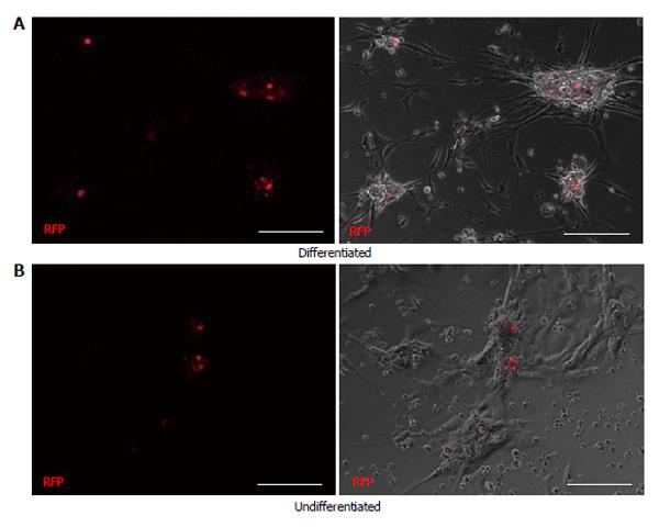
Figure 3 Comparison of differentiated (A) and undifferentiated (B) cells from the islet depleted pancreatic tissue infected with PDX1-monomeric red fluorescent protein.
A few PDX1+ cells (RFP) are visible within the adhered aggregates in the differentiated condition (A). Cell aggregates are absent in the undifferentiated cell conditions (B) and PDX1+ cells are within the monolayer. Scale bars are 100 μm. RFP: Red fluorescent protein.
- Citation: Seeberger KL, Anderson SJ, Ellis CE, Yeung TY, Korbutt GS. Identification and differentiation of PDX1 β-cell progenitors within the human pancreatic epithelium. World J Diabetes 2014; 5(1): 59-68
- URL: https://www.wjgnet.com/1948-9358/full/v5/i1/59.htm
- DOI: https://dx.doi.org/10.4239/wjd.v5.i1.59


