Copyright
©2013 Baishideng Publishing Group Co.
World J Diabetes. Dec 15, 2013; 4(6): 310-318
Published online Dec 15, 2013. doi: 10.4239/wjd.v4.i6.310
Published online Dec 15, 2013. doi: 10.4239/wjd.v4.i6.310
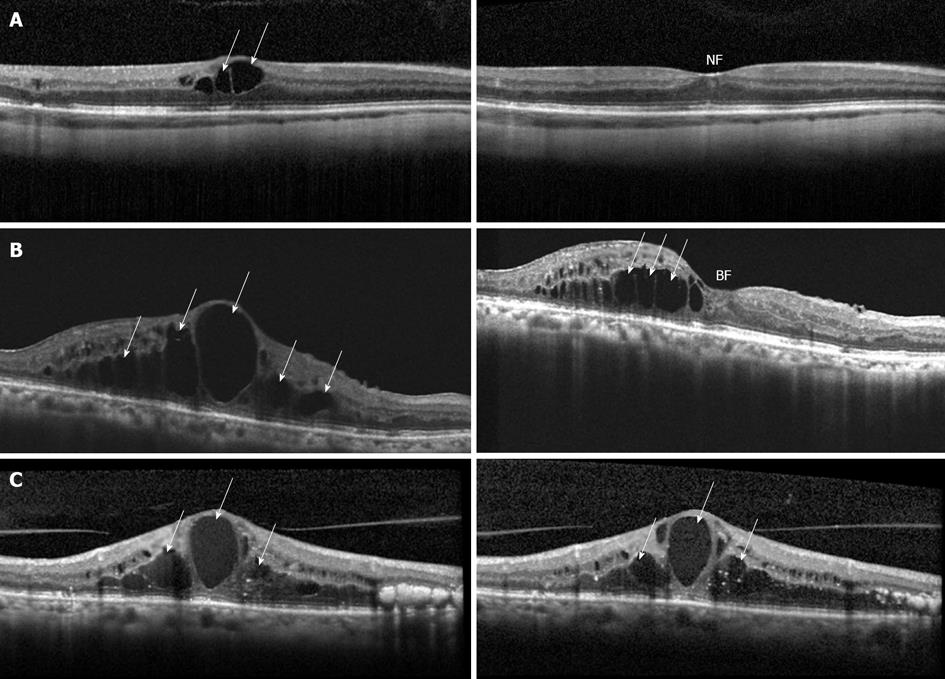
Figure 3 High resolution optical coherence tomography demonstrating different responses to treatment of diabetic macular edema patients with ranibizumab.
A: Modest cystoid macular edema (CME) with few inner retinal cysts (white arrows) and loss of the foveal contour (left) which completely resolved with return of a normal foveal contour (NF) and excellent vision one month after a single injection of ranibizumab (right); B: Massive CME with numerous inner and outer retinal cysts (white arrows) with complete loss of the foveal contour (left) which partially resolves resulting in a blunted but improved foveal contour (BF) and a significant improvement in vision following treatment with ranibizumab (right); C: Massive CME with numerous inner and outer retinal cysts (white arrows) with complete loss of the foveal contour (left) which does not respond despite repeated treatment with ranibizumab (right). The last response is uncommon; this patient was ultimately treated with intraocular steroids, and did have a sustained improvement of the edema and a modest improvement in vision.
- Citation: Krispel C, Rodrigues M, Xin X, Sodhi A. Ranibizumab in diabetic macular edema. World J Diabetes 2013; 4(6): 310-318
- URL: https://www.wjgnet.com/1948-9358/full/v4/i6/310.htm
- DOI: https://dx.doi.org/10.4239/wjd.v4.i6.310


