Copyright
©2013 Baishideng Publishing Group Co.
World J Diabetes. Apr 15, 2013; 4(2): 19-26
Published online Apr 15, 2013. doi: 10.4239/wjd.v4.i2.19
Published online Apr 15, 2013. doi: 10.4239/wjd.v4.i2.19
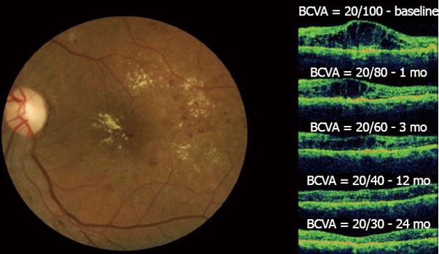
Figure 5 Diffuse diabetic macular edema treated with bevacizumab.
In the left figure, the clinical fundus photograph shows the macular edema and hard exudates at the foveal center.In the right figure, a series of optical coherence tomographys (OCTs) taken at a 24-mo follow-up can be observed. The OCT image at baseline shows the intraretinal fluid with increased central macular thickness (CMT) and best-corrected visual acuity (BCVA) = 20/100. One month after the first injection, improvement in both BCVA and CMT was observed. This result was maintained throughout the 24-mo follow-up period after six injections and with final central macular thickness within normal limits without intraretinal fluid and the improvement of BCVA to 20/30. No laser photocoagulation was performed in this case.
- Citation: Stefanini FR, Arevalo JF, Maia M. Bevacizumab for the management of diabetic macular edema. World J Diabetes 2013; 4(2): 19-26
- URL: https://www.wjgnet.com/1948-9358/full/v4/i2/19.htm
- DOI: https://dx.doi.org/10.4239/wjd.v4.i2.19


