Copyright
©2013 Baishideng Publishing Group Co.
World J Diabetes. Apr 15, 2013; 4(2): 19-26
Published online Apr 15, 2013. doi: 10.4239/wjd.v4.i2.19
Published online Apr 15, 2013. doi: 10.4239/wjd.v4.i2.19
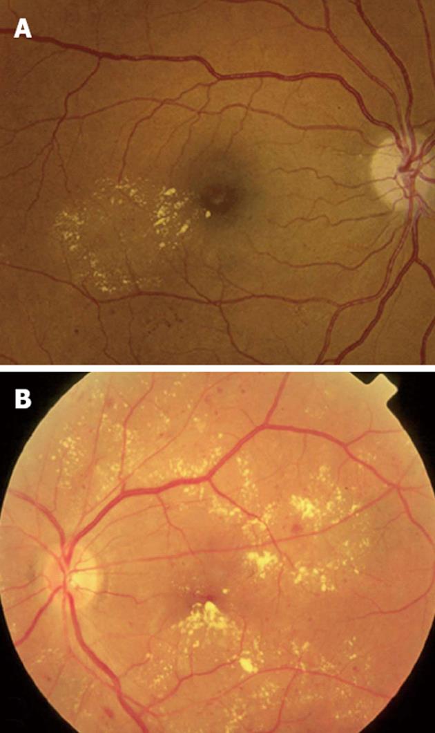
Figure 2 Clinical patterns of diabetic macular edema.
A: Focal macular edema marked by focal leakage from microaneurysms and dilated retinal capillaries with abnormal permeability, making a complete ring as a localized circinate pattern of hard exudates; B: Diffuse macular edema, characterized by hard exudates with generalized leakage from dilated capillaries throughout the posterior pole.
- Citation: Stefanini FR, Arevalo JF, Maia M. Bevacizumab for the management of diabetic macular edema. World J Diabetes 2013; 4(2): 19-26
- URL: https://www.wjgnet.com/1948-9358/full/v4/i2/19.htm
- DOI: https://dx.doi.org/10.4239/wjd.v4.i2.19


