Copyright
©The Author(s) 2025.
World J Diabetes. May 15, 2025; 16(5): 101473
Published online May 15, 2025. doi: 10.4239/wjd.v16.i5.101473
Published online May 15, 2025. doi: 10.4239/wjd.v16.i5.101473
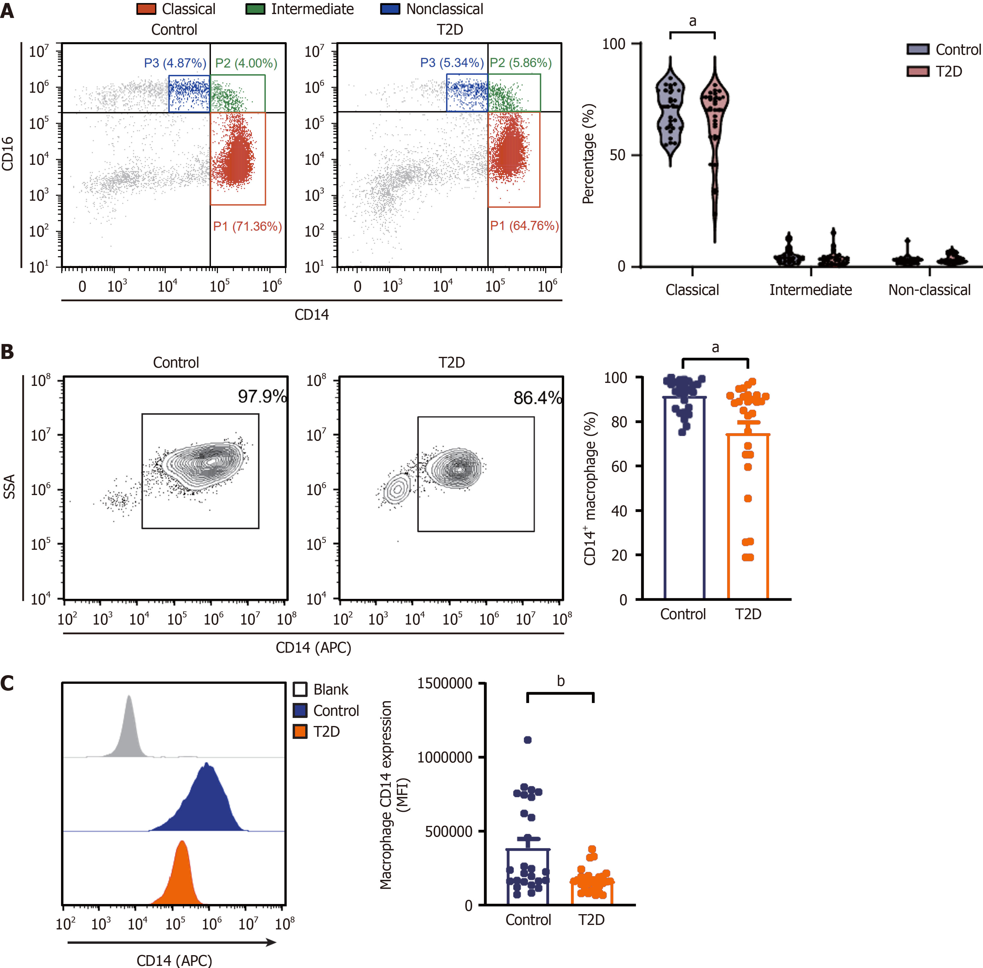
Figure 2 Percentage of classical monocytes and macrophage CD14 expression were decreased in type 2 diabetes patients.
A: Percentages of different monocyte subsets (classical, intermediate and nonclassical monocytes); B: The percentage of CD14+ macrophages in control subjects and diabetes patients was determined by flow cytometry; C: The median fluorescence intensity of CD14 in macrophages from control subjects and diabetes patients. The data are the mean ± SD. aP < 0.05, bP < 0.01 from paired t test. T2D: Type 2 diabetes; MFI: Median fluorescence intensity.
- Citation: Mao QY, Ran H, Hu QY, He SY, Lu Y, Li H, Chai YM, Chu ZY, Qian X, Ding W, Niu YX, Zhang HM, Li XY, Su Q. Impaired efferocytosis by monocytes and monocyte-derived macrophages in patients with poorly controlled type 2 diabetes. World J Diabetes 2025; 16(5): 101473
- URL: https://www.wjgnet.com/1948-9358/full/v16/i5/101473.htm
- DOI: https://dx.doi.org/10.4239/wjd.v16.i5.101473


