Copyright
©The Author(s) 2025.
World J Diabetes. Mar 15, 2025; 16(3): 97336
Published online Mar 15, 2025. doi: 10.4239/wjd.v16.i3.97336
Published online Mar 15, 2025. doi: 10.4239/wjd.v16.i3.97336
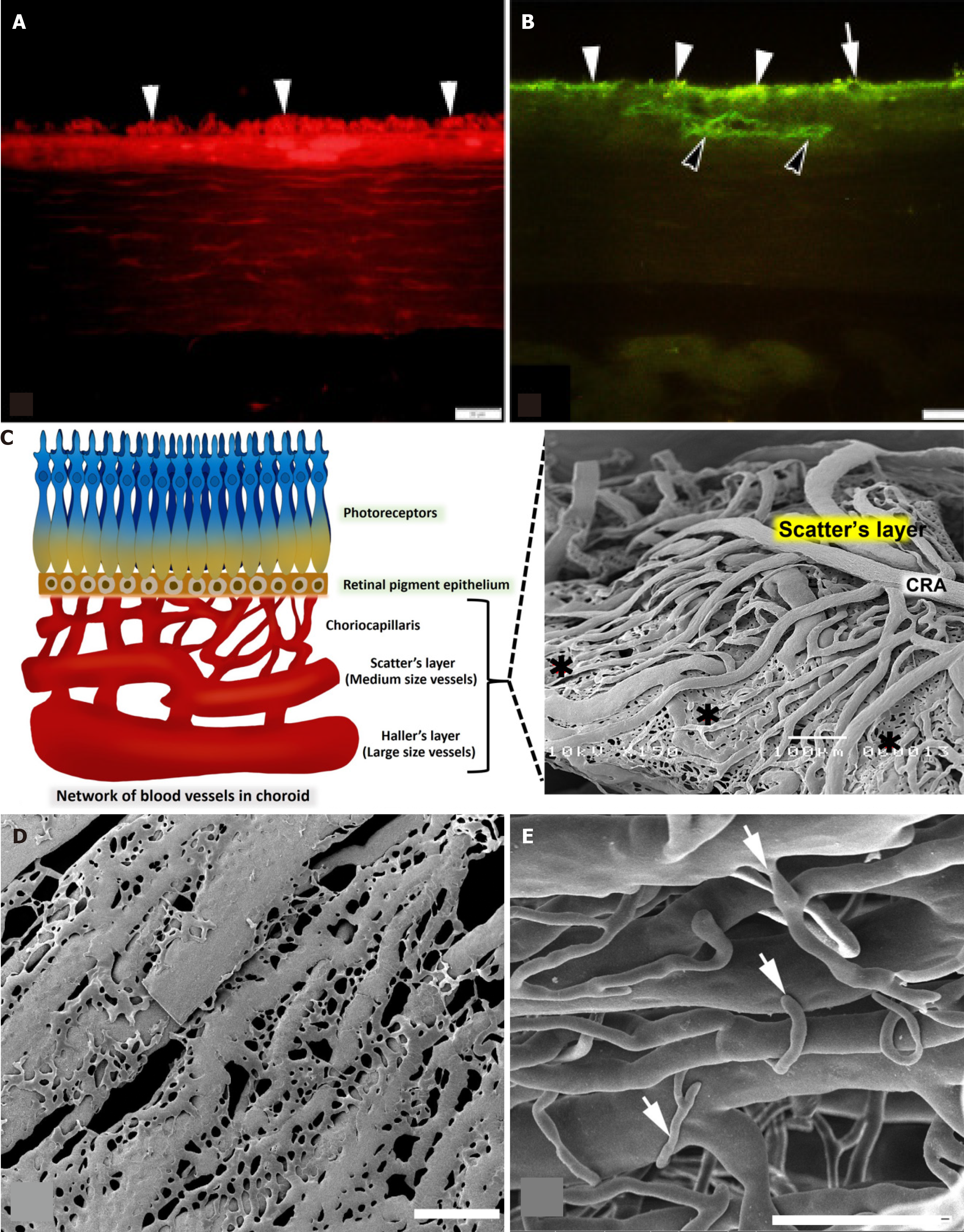
Figure 6 Schematic diagrams and the representation of results.
A: Photomicrographs of immunohistochemical sections of the choroidal layer with specific antibodies in the diabetic group. An example of immunoreactive vascular endothelial growth factor in the choriocapillaris and the retinal pigment epithelium (RPE) (white arrowheads); B: Immunoreactive cluster of differentiation 31 in the region of the RPE (white arrowheads), choriocapillaris (white arrow), and the wall of blood vessels (black arrowheads) was present in diabetic group. Scale bar = 20 μm; C: A comparison of the normal choroidal vascular structure and the vascular corrosion casting framework is indicated in this diagram. The outermost layer of the choroid, which is also known as Haller’s layer, is composed of vessels with a large vessel capacity. Sattler’s layer is the name given to the innermost layer of the choroid, which is composed of vessels that are significantly smaller in size than the vessels that make up the outermost layer such as choroidal artery. An extensive number of anastomotic capillaries are what constitute the choriocapillaris (black roundness) of the choroid; D: Scanning electron microscopy (SEM) micrographs using a vascular corrosion casting technique/SEM in the diabetic group. The arrangement of the choriocapillaris is not dense and untidy; they link to one another, resulting in the formation of a flat sheet; E: Furthermore, the sprouting of blood vessels (white arrows) was discovered in Sattler’s layer, which included choroidal arteries that were significantly more exposed. Scale bar = 300 μm. CRA: Choroid arteries.
- Citation: Matsathit U, Komolkriengkrai M, Khimmaktong W. Glabridin and gymnemic acid alleviates choroid structural change and choriocapillaris impairment in diabetic rat’s eyes. World J Diabetes 2025; 16(3): 97336
- URL: https://www.wjgnet.com/1948-9358/full/v16/i3/97336.htm
- DOI: https://dx.doi.org/10.4239/wjd.v16.i3.97336


