Copyright
©The Author(s) 2025.
World J Diabetes. Mar 15, 2025; 16(3): 102277
Published online Mar 15, 2025. doi: 10.4239/wjd.v16.i3.102277
Published online Mar 15, 2025. doi: 10.4239/wjd.v16.i3.102277
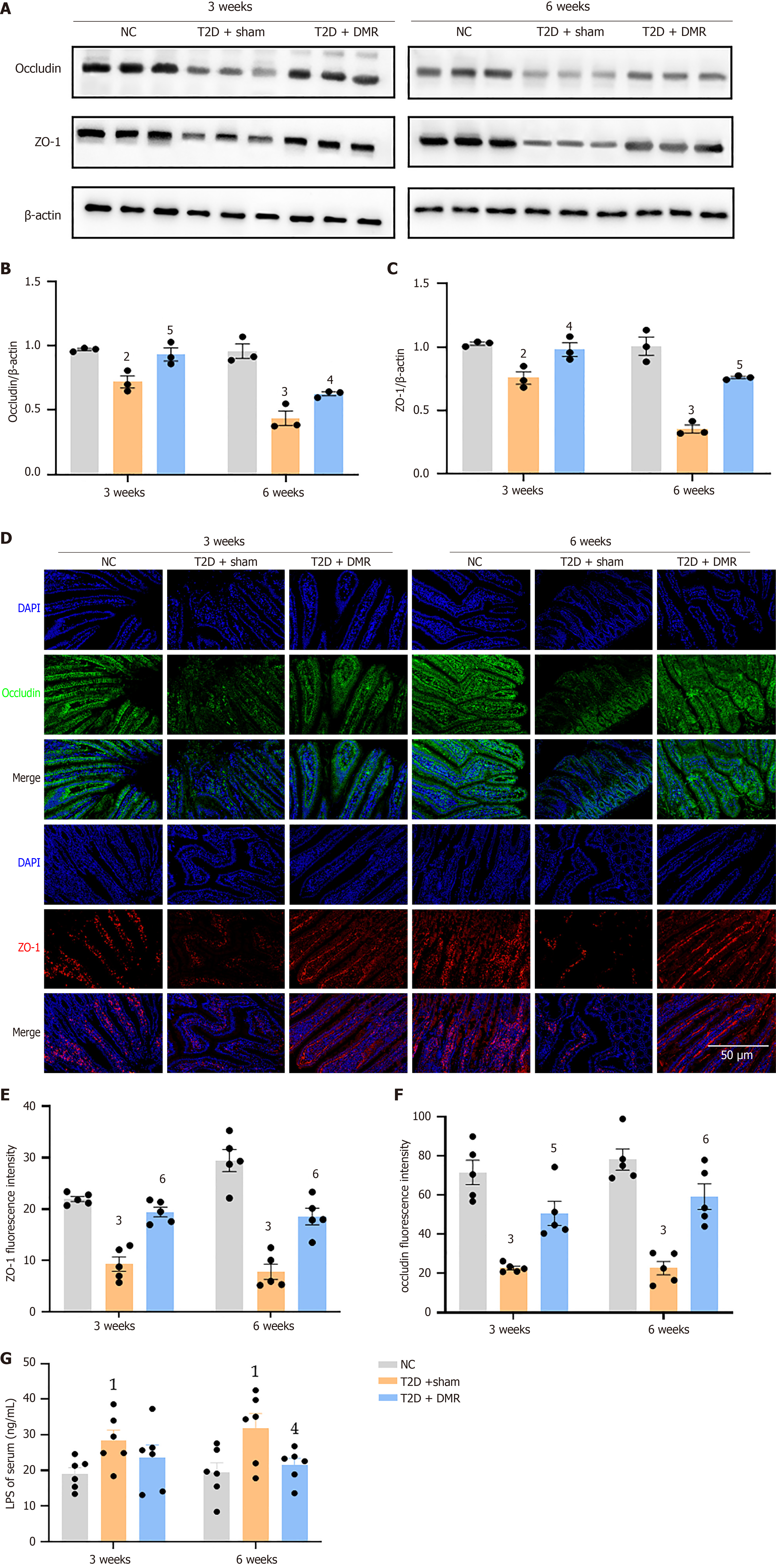
Figure 4 Effects of the duodenal mucosal resurfacing procedure on the duodenal mucosal barrier function in type 2 diabetes rats.
A: Protein extracts of duodenum were subjected to western blot analysis with antibodies to occludin, zonula occludens-1 (ZO-1) or β-actin; B and C: The blots of occludin and ZO-1 were quantified using image J software and normalized to β-actin levels (n = 3 mice per group); D: Representative images of immunofluorescence staining for occludin (green), ZO-1 (red) and 4’,6-diamidino-2-phenylindole (blue) of duodenum (Scale bar = 50 μm); E and F: The relative fluorescence intensity of occludin and ZO-1 (n = 5 mice per group); G: Serum lipopolysaccharide levels of rats at postoperative weeks 3 and 6 (n = 6 mice per group). Data are presented as mean ± SE. 1P < 0.05 vs normal control group. 2P < 0.01 vs normal control group. 3P < 0.001 vs normal control group. 4P < 0.05 vs type 2 diabetes-sham group. 5P < 0.01 vs type 2 diabetes-sham group. 6P < 0.001 vs type 2 diabetes-sham group. DMR: Duodenal mucosal resurfacing; T2D: Type 2 diabetes; NC: Normal control; LPS: Lipopolysaccharide; ZO-1: Zonula occludens-1; DAPI: 4’,6-diamidino-2-phenylindole.
- Citation: Nie LJ, Cheng Z, He YX, Yan QH, Sun YH, Yang XY, Tian J, Zhu PF, Yu JY, Zhou HP, Zhou XQ. Role of duodenal mucosal resurfacing in controlling diabetes in rats. World J Diabetes 2025; 16(3): 102277
- URL: https://www.wjgnet.com/1948-9358/full/v16/i3/102277.htm
- DOI: https://dx.doi.org/10.4239/wjd.v16.i3.102277


