Copyright
©The Author(s) 2025.
World J Diabetes. Mar 15, 2025; 16(3): 101328
Published online Mar 15, 2025. doi: 10.4239/wjd.v16.i3.101328
Published online Mar 15, 2025. doi: 10.4239/wjd.v16.i3.101328
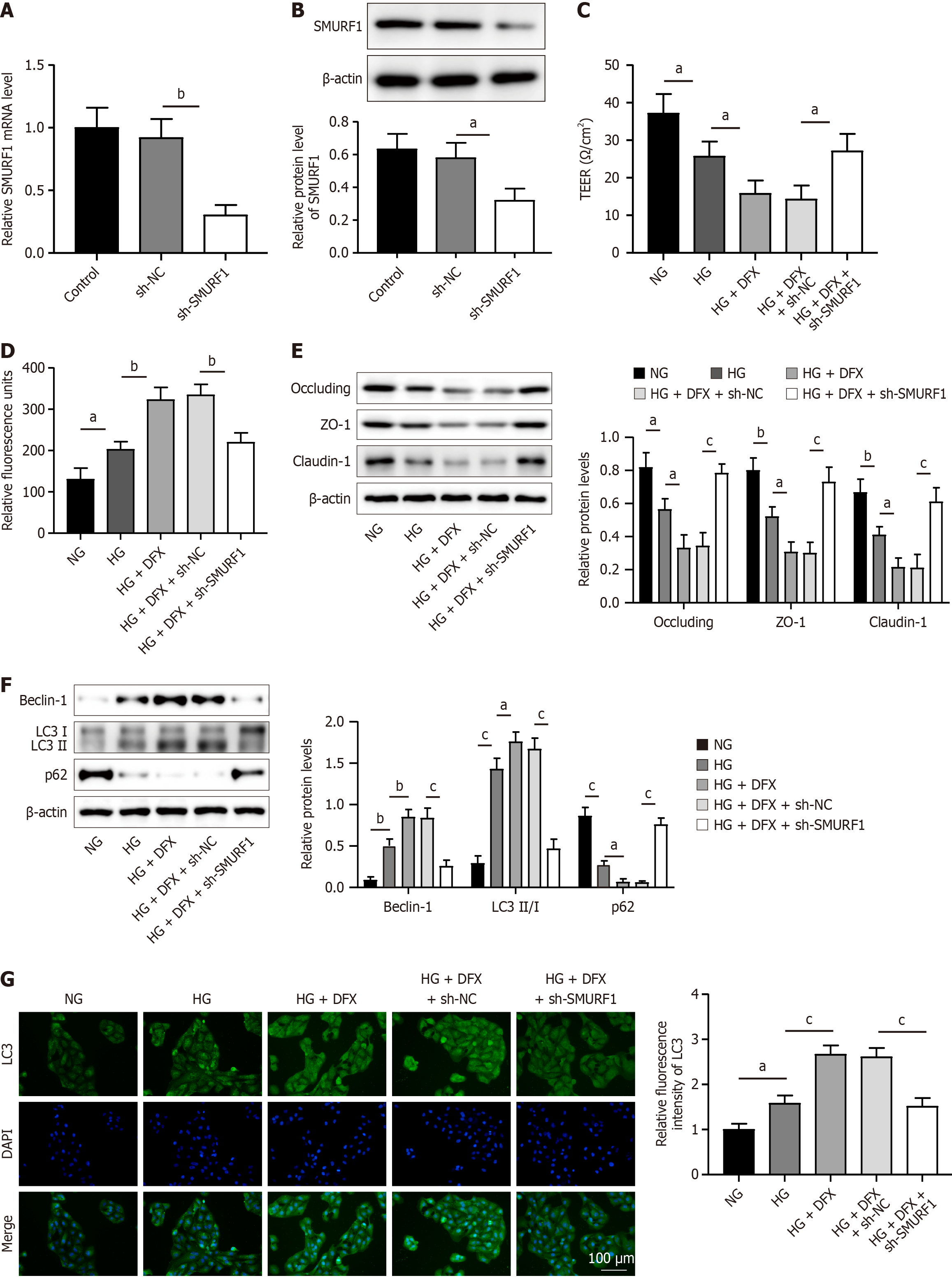
Figure 2 SMAD-specific E3 ubiquitin protein ligase 1 knockdown promoted retinal pigment epithelium cell tight junctions by inhibiting autophagy in diabetic macular edema.
A: The SMAD-specific E3 ubiquitin protein ligase (SMURF) 1 knockdown construct or vector were transfected in ARPE-19 cells, and the mRNA level of SMURF1 was then detected by quantitative real-time PCR; B: ARPE-19 cells transfected with the vector or SMURF1 construct were collected, followed by western blot to detect SMURF1 protein levels; C: Vector or SMURF1 knockdown ARPE-19 cells were treated with normal concentration of glucose (NG), high concentration of glucose (HG) or desferrioxamine mesylate (DFX), followed by determination of cellular trans-epithelial electrical resistance; D: Vector or SMURF1 knockdown ARPE-19 cells were treated with NG, HG or DFX, and the detection of cell permeability was performed; E: Vector or SMURF1 knockdown ARPE-19 cells were treated with NG, HG or DFX, and then the expression of occluding, ZO-1, and claudin-1 was detected by western blot; F: Vector or SMURF1 knockdown ARPE-19 cells were treated with NG, HG or DFX, and the expression of Beclin-1, LC3I, LC3II, and p62 was then detected by western blot; G: Vector or SMURF1 knockdown ARPE-19 cells were treated with NG, HG or DFX, followed by immunofluorescence analysis of LC3. The experiments were repeated three times. aP < 0.05, bP < 0.01, cP < 0.001. SMURF1: SMAD-specific E3 ubiquitin protein ligase 1; NG: Normal concentration of glucose; HG: High concentration of glucose; DFX: Desferrioxamine mesylate.
- Citation: Liang LF, Zhao JQ, Wu YF, Chen HJ, Huang T, Lu XH. SMAD specific E3 ubiquitin protein ligase 1 accelerates diabetic macular edema progression by WNT inhibitory factor 1. World J Diabetes 2025; 16(3): 101328
- URL: https://www.wjgnet.com/1948-9358/full/v16/i3/101328.htm
- DOI: https://dx.doi.org/10.4239/wjd.v16.i3.101328


