Copyright
©The Author(s) 2025.
World J Diabetes. Feb 15, 2025; 16(2): 94861
Published online Feb 15, 2025. doi: 10.4239/wjd.v16.i2.94861
Published online Feb 15, 2025. doi: 10.4239/wjd.v16.i2.94861
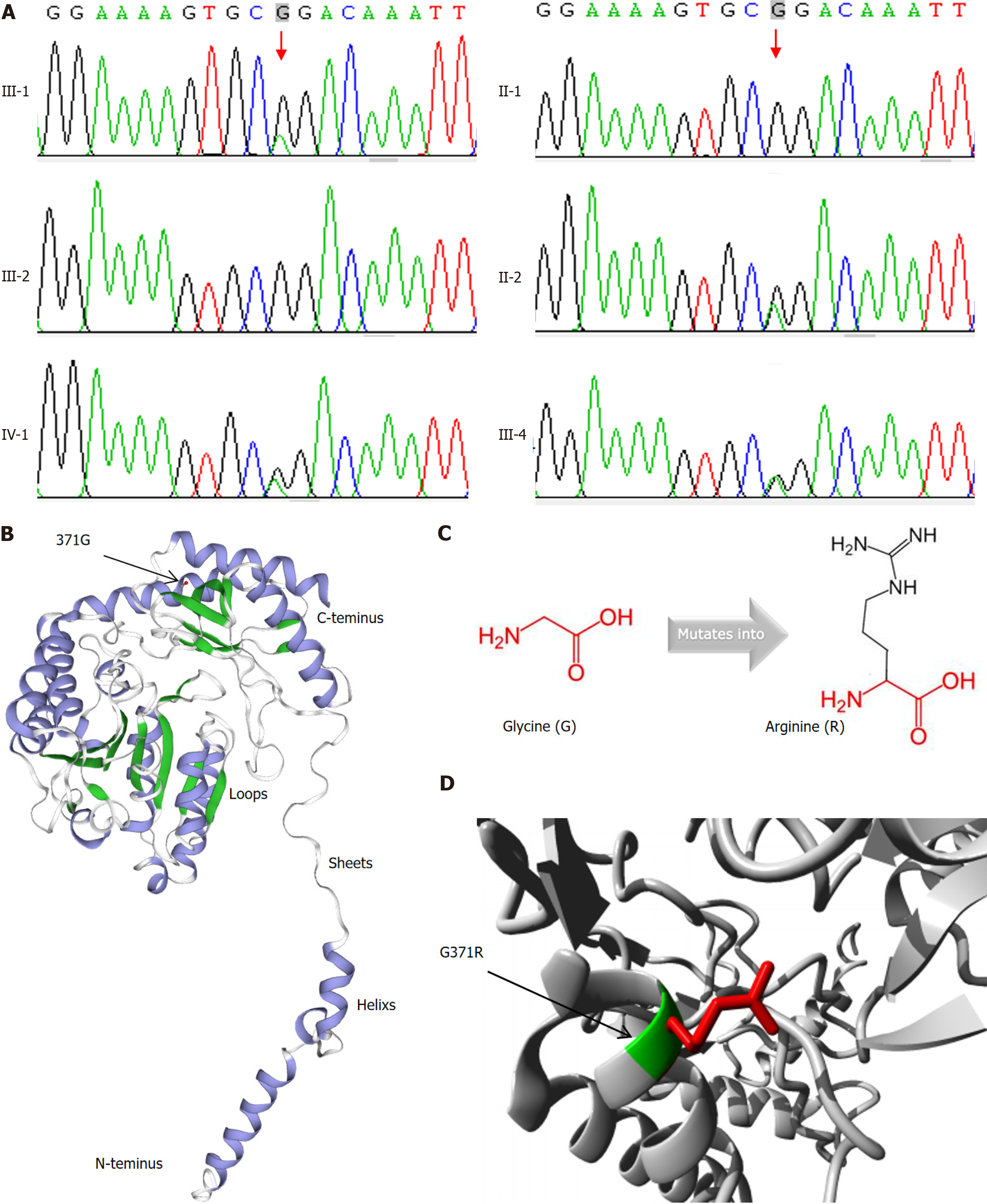
Figure 3 Description of the mutation.
A: The sanger sequence analysis of p.G371R mutation of SPTLC1 gene; B: Three dimensional (3D) structures of wild-type of human SPTLC1 gene; C: Structural formulas show that the mutant residue (R) is bigger than the wild-type residue (G); D: Zoomed 3D structure of mutation of human SPTLC1.
- Citation: Yi B, Bao Y, Wen ZY. Effect of SPTLC1 on type 2 diabetes mellitus. World J Diabetes 2025; 16(2): 94861
- URL: https://www.wjgnet.com/1948-9358/full/v16/i2/94861.htm
- DOI: https://dx.doi.org/10.4239/wjd.v16.i2.94861


