Copyright
©The Author(s) 2024.
World J Diabetes. Sep 15, 2024; 15(9): 1979-2001
Published online Sep 15, 2024. doi: 10.4239/wjd.v15.i9.1979
Published online Sep 15, 2024. doi: 10.4239/wjd.v15.i9.1979
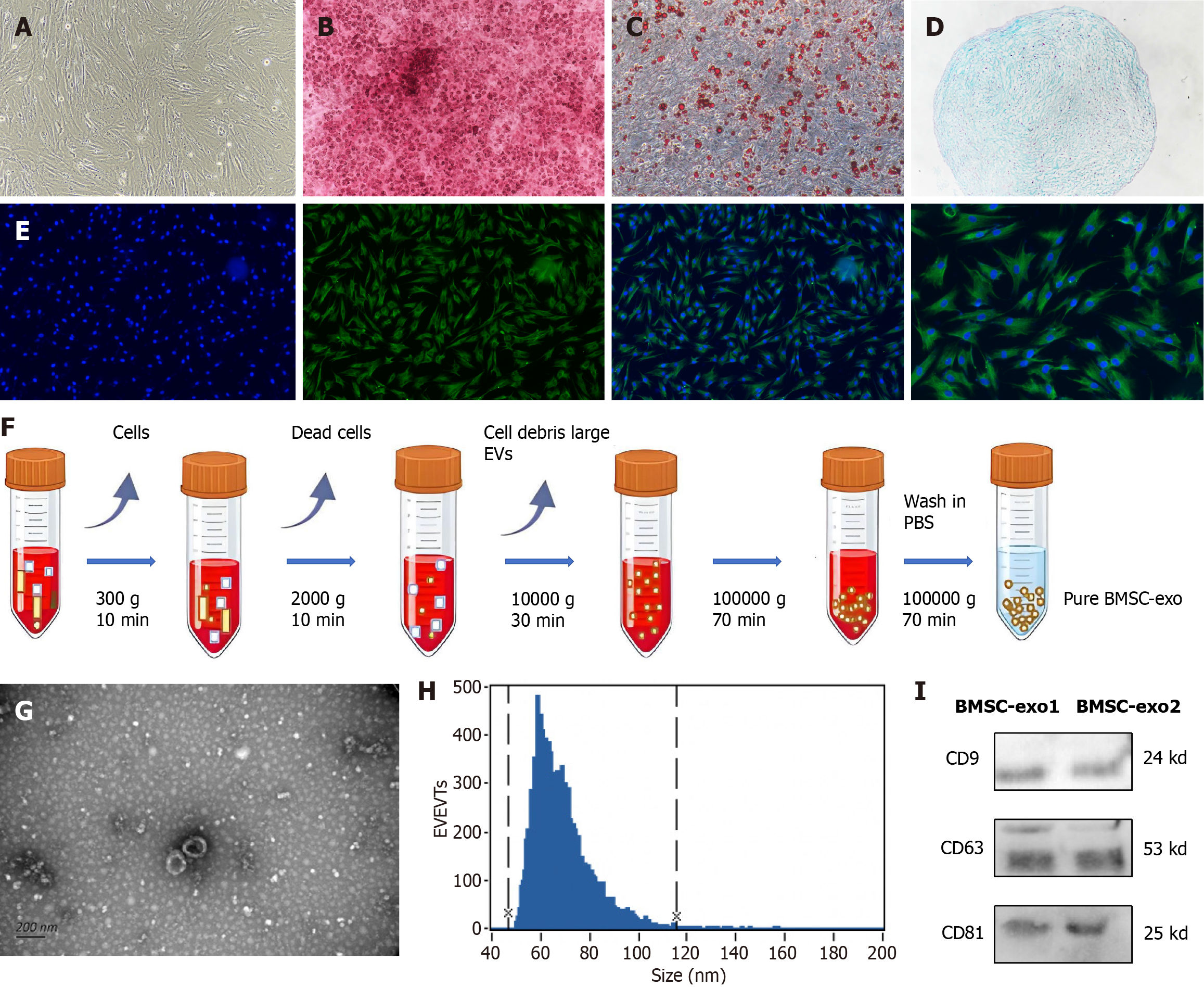
Figure 2 Characterization of bone marrow-derived mesenchymal stem cells and bone marrow-derived mesenchymal stem cell-derived exosomes.
A: Bone marrow-derived mesenchymal stem cells (BMSC) were fusiform. Scale = 50 μm; B: Osteoblast alizarin red, before and after staining. Scale = 50 μm; C: Lipoblast oil red O, before and after staining. Scale = 50 μm; D: Formation of chondrospheres, stained with alcian blue. Scale = 50 μm; E: Cluster of differentiation (CD) 44 immunofluorescence staining. Scale = 50 μm/25 μm; F: Extraction of BMSC-derived exosomes (BMSC-exo) by ultrafast centrifugation; G: BMSC-exo appeared saucer-shaped under electron microscopy. Scale = 200 nm; H: BMSC-exo particle microscopy with 47 nm and 116 nm diameters; I: Western blot analysis of protein expression of exosome markers: CD9; CD63; and CD81. EVs: Extracellular vesicles; PBS: Phosphate buffered saline; BMSC-exo: Bone marrow mesenchymal stem cell derived exosomes.
- Citation: Wang YY, Li K, Wang JJ, Hua W, Liu Q, Sun YL, Qi JP, Song YJ. Bone marrow-derived mesenchymal stem cell-derived exosome-loaded miR-129-5p targets high-mobility group box 1 attenuates neurological-impairment after diabetic cerebral hemorrhage. World J Diabetes 2024; 15(9): 1979-2001
- URL: https://www.wjgnet.com/1948-9358/full/v15/i9/1979.htm
- DOI: https://dx.doi.org/10.4239/wjd.v15.i9.1979


