Copyright
©The Author(s) 2024.
World J Diabetes. Jul 15, 2024; 15(7): 1499-1508
Published online Jul 15, 2024. doi: 10.4239/wjd.v15.i7.1499
Published online Jul 15, 2024. doi: 10.4239/wjd.v15.i7.1499
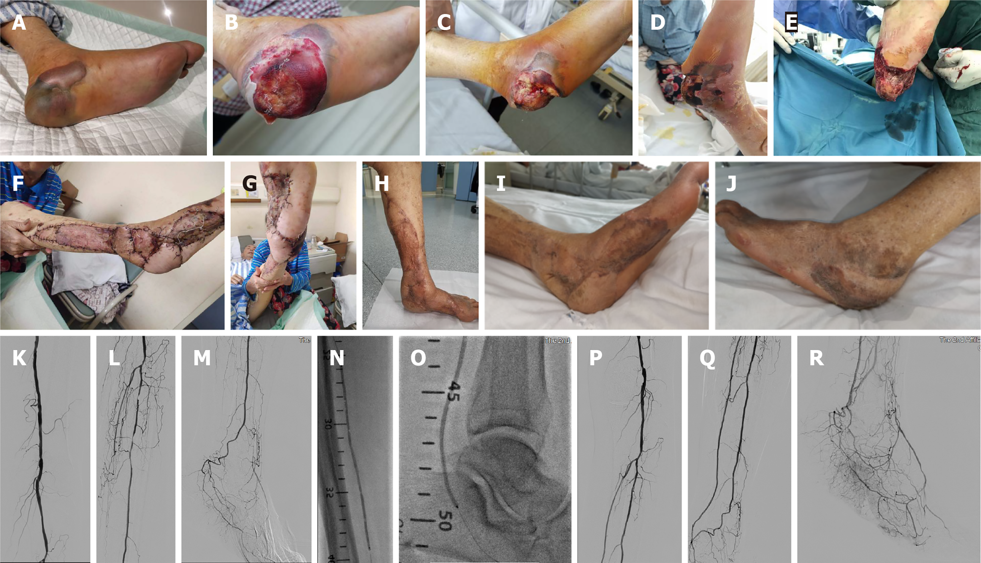
Figure 4 Surgery pictures of a local infection case.
A-D: The heel of a diabetic foot patient was punctured by a nail, resulting in local infection and gradual degeneration and necrosis of the soft tissue of the heel; E-G: Complete debridement was performed after revascularization, and the heel was repaired with a free-style perforator flap on the lower leg side; H-J: Two months after surgery, the patient's foot was successfully preserved and functioned well; K-M: Angiography showed patency of the femoral-popliteal artery, occlusions of the infrapopliteal and anterior tibial veins, occlusion of the middle and distal posterior tibial artery, and staged stenosis of peroneal artery; N and O: balloon angioplasty (BA) of posterior tibial artery and peroneal artery was performed; P-R: Angiography after BA showed that the blood flow of posterior tibial artery and peroneal artery was smooth with the blood flow directly to the heel, and the plantar arterial arch was good.
- Citation: Lei FR, Shen XF, Zhang C, Li XQ, Zhuang H, Sang HF. Clinical efficacy of endovascular revascularization combined with vacuum-assisted closure for the treatment of diabetic foot. World J Diabetes 2024; 15(7): 1499-1508
- URL: https://www.wjgnet.com/1948-9358/full/v15/i7/1499.htm
- DOI: https://dx.doi.org/10.4239/wjd.v15.i7.1499


