Copyright
©The Author(s) 2024.
World J Diabetes. Jul 15, 2024; 15(7): 1499-1508
Published online Jul 15, 2024. doi: 10.4239/wjd.v15.i7.1499
Published online Jul 15, 2024. doi: 10.4239/wjd.v15.i7.1499
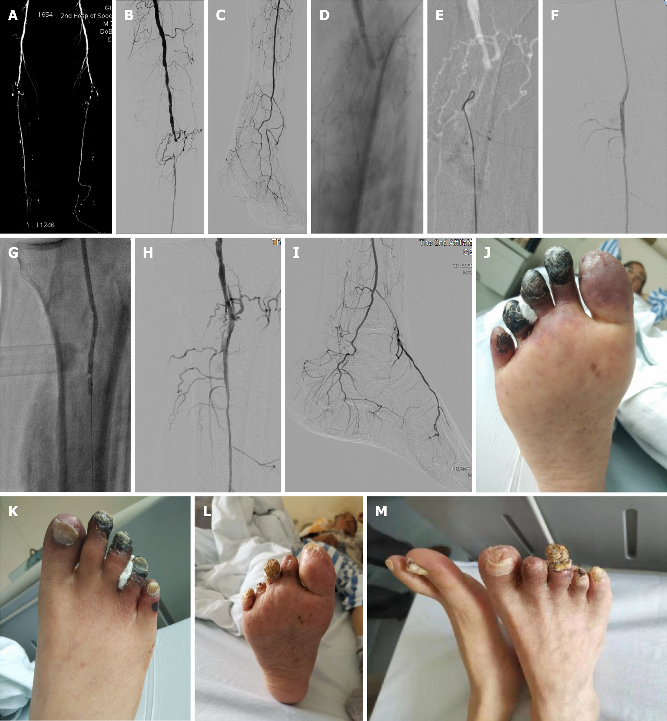
Figure 3 Surgery pictures of a diabetic foot complicated with uremia.
A: Lower limb computerized tomography angiography of a diabetic foot complicated with uremia showed proximal occlusion of the peroneal artery, the only outflow tract below the knee, obvious arterial calcification; B-D: Angiography showed that the proximal peroneal artery was occluded, and the guidewire could not pass through the lesion due to severe calcification; E-G: Retrograde puncture of the distal peroneal artery was performed, and balloon angioplasty (BA) was carried out after the orbit was established by connecting the guidewire with the proximal catheter; H and I: The blood flow was obviously improved after BA with non-flow-limiting dissection locally visible, and no stent was implanted; J: Gangrene at the ends of the 2nd, 3rd and 4th toes of the left foot; K: Cyanosis at the 1st and 5th toes; L and M: Four weeks later, the necrotic part of the toe fell off spontaneously and the wound healed spontaneously.
- Citation: Lei FR, Shen XF, Zhang C, Li XQ, Zhuang H, Sang HF. Clinical efficacy of endovascular revascularization combined with vacuum-assisted closure for the treatment of diabetic foot. World J Diabetes 2024; 15(7): 1499-1508
- URL: https://www.wjgnet.com/1948-9358/full/v15/i7/1499.htm
- DOI: https://dx.doi.org/10.4239/wjd.v15.i7.1499


