Copyright
©The Author(s) 2024.
World J Diabetes. Jun 15, 2024; 15(6): 1299-1316
Published online Jun 15, 2024. doi: 10.4239/wjd.v15.i6.1299
Published online Jun 15, 2024. doi: 10.4239/wjd.v15.i6.1299
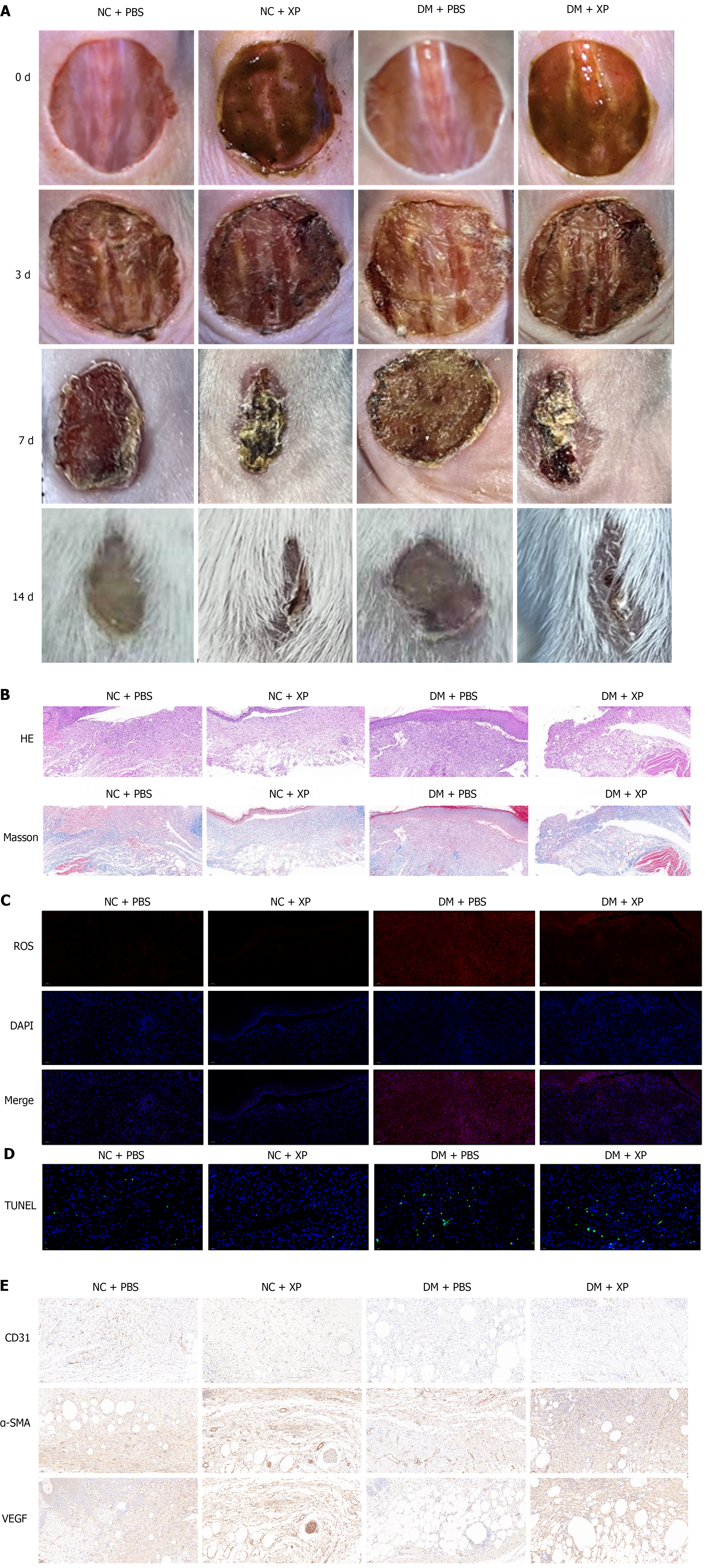
Figure 1 X-Paste promotes wound healing in diabetic foot ulcers mice.
A: Images of mice grouped in either normal control (NC), NC plus X-Paste (XP), diabetic wound plus phosphate-buffered saline or diabetic wound plus XP were captured at days 0, 3, 7 and 14; B: Wound tissue sections were stained with Hematoxylin and Eosin and Masson in different groups; C: The representative images of reactive oxygen species production in wound tissue sections from different groups; D: The representative images of TUNEL in wound tissue sections from different groups; E: The representative images of CD31, alpha-smooth muscle actin, and VEGFA from different groups. NC: Normal control; PBS: Phosphate-buffered saline; DM: Diabetic wound; XP: X-Paste; α-SMA: Alpha-smooth muscle actin.
- Citation: Du MW, Zhu XL, Zhang DX, Chen XZ, Yang LH, Xiao JZ, Fang WJ, Xue XC, Pan WH, Liao WQ, Yang T. X-Paste improves wound healing in diabetes via NF-E2-related factor/HO-1 signaling pathway. World J Diabetes 2024; 15(6): 1299-1316
- URL: https://www.wjgnet.com/1948-9358/full/v15/i6/1299.htm
- DOI: https://dx.doi.org/10.4239/wjd.v15.i6.1299


