Copyright
©The Author(s) 2024.
World J Diabetes. Jun 15, 2024; 15(6): 1280-1290
Published online Jun 15, 2024. doi: 10.4239/wjd.v15.i6.1280
Published online Jun 15, 2024. doi: 10.4239/wjd.v15.i6.1280
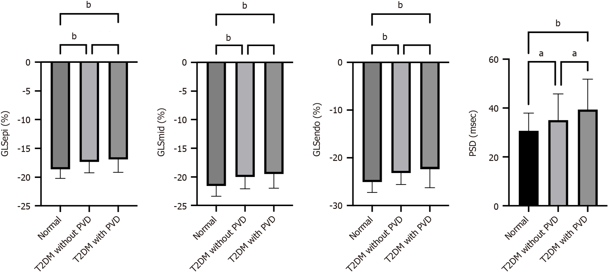
Figure 1 Line graph showing the differences in layer-specific global longitudinal strain and peak strain dispersion between normal controls, type 2 diabetes mellitus patients without peripheral vascular disease and type 2 diabetes mellitus patients with peripheral vascular disease.
aP < 0.05; bP < 0.01. GLSEpi: Global longitudinal strain of the epimyocardium; GLSMid: Global longitudinal strain of the middle myocardium; GLSEndo: Global longitudinal strain of the endocardium; PSD: Peak strain dispersion; LDL-C: Low-density lipoprotein cholesterol; LVEF: Left ventricular ejection fraction; PVD: Peripheral vascular disease; T2DM: Type 2 diabetes mellitus.
- Citation: Li GA, Huang J, Fan L. Evaluation of left ventricular systolic function in type 2 diabetes mellitus patients with and without peripheral vascular disease. World J Diabetes 2024; 15(6): 1280-1290
- URL: https://www.wjgnet.com/1948-9358/full/v15/i6/1280.htm
- DOI: https://dx.doi.org/10.4239/wjd.v15.i6.1280


