Copyright
©The Author(s) 2024.
World J Diabetes. Apr 15, 2024; 15(4): 769-782
Published online Apr 15, 2024. doi: 10.4239/wjd.v15.i4.769
Published online Apr 15, 2024. doi: 10.4239/wjd.v15.i4.769
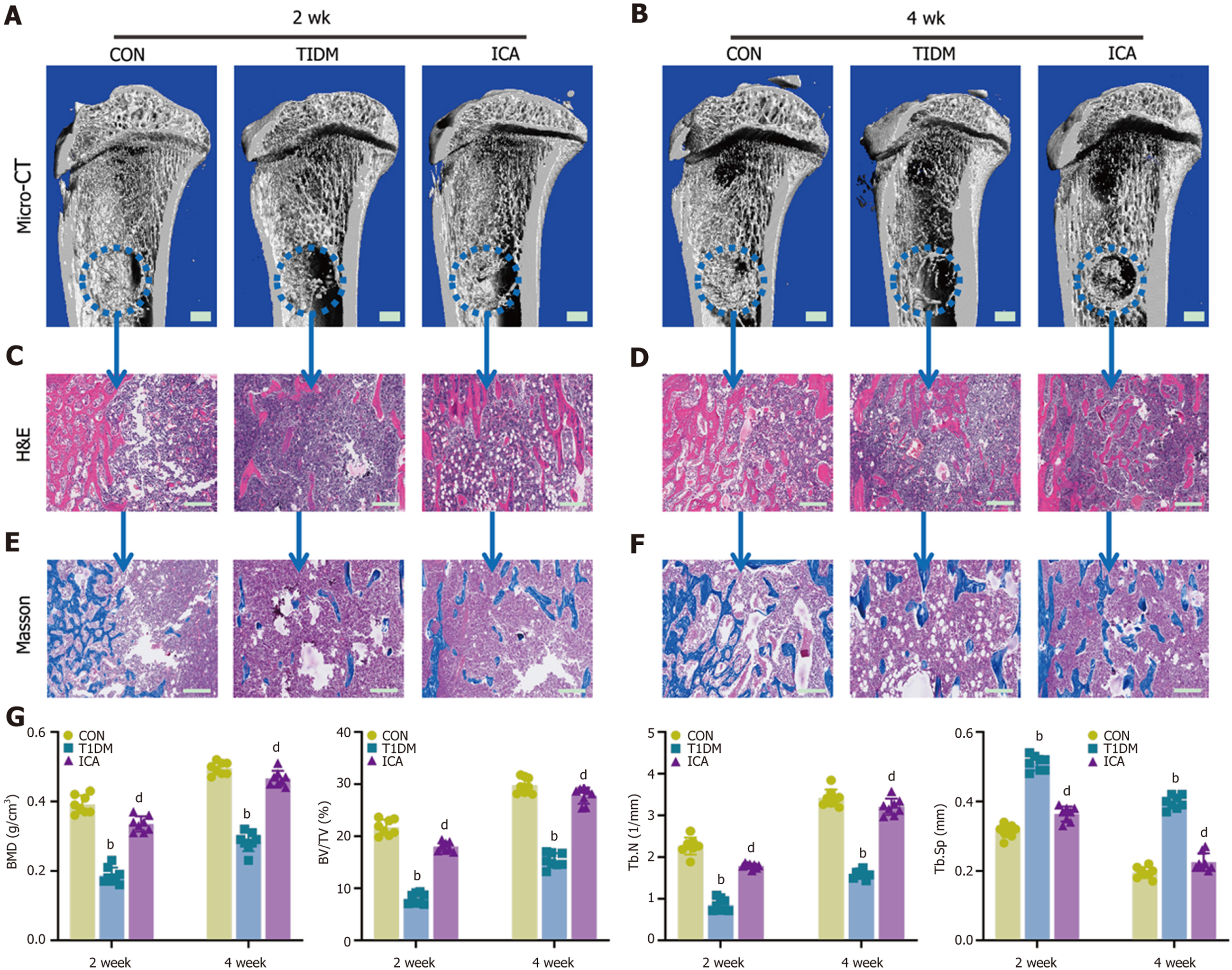
Figure 5 Icariin improved bone repair capacity by promoting osteogenesis in type 1 diabetes mellitus rats.
A and B: Three-dimensional reconstruction images of the defect area at week 2 (A) and week 4 (B) (scale bars: 1 mm); C and D: Hematoxylin and eosin staining of the defect area at week 2 (C) and week 4 (B) (scale bars: 200 μm); E and F: Masson’s trichrome staining of the defect area at week 2 (E) and week 4 (F) (scale bars: 200 μm); G: Micron-scale computed tomography analysis of bone mineral density (BMD), bone volume/tissue volume (BV/TV), trabecular number (Tb.N), and trabecular separation (Tb.Sp) at weeks 2 and 4. Data are mean ± SEM of the mean (n = 8). bP < 0.01 vs the control group; dP < 0.01 vs the type 1 diabetes mellitus group. ICA: Icariin; Con: Control; T1DM: Type 1 diabetes mellitus.
- Citation: Zheng S, Hu GY, Li JH, Zheng J, Li YK. Icariin accelerates bone regeneration by inducing osteogenesis-angiogenesis coupling in rats with type 1 diabetes mellitus. World J Diabetes 2024; 15(4): 769-782
- URL: https://www.wjgnet.com/1948-9358/full/v15/i4/769.htm
- DOI: https://dx.doi.org/10.4239/wjd.v15.i4.769


