Copyright
©The Author(s) 2024.
World J Diabetes. Dec 15, 2024; 15(12): 2322-2337
Published online Dec 15, 2024. doi: 10.4239/wjd.v15.i12.2322
Published online Dec 15, 2024. doi: 10.4239/wjd.v15.i12.2322
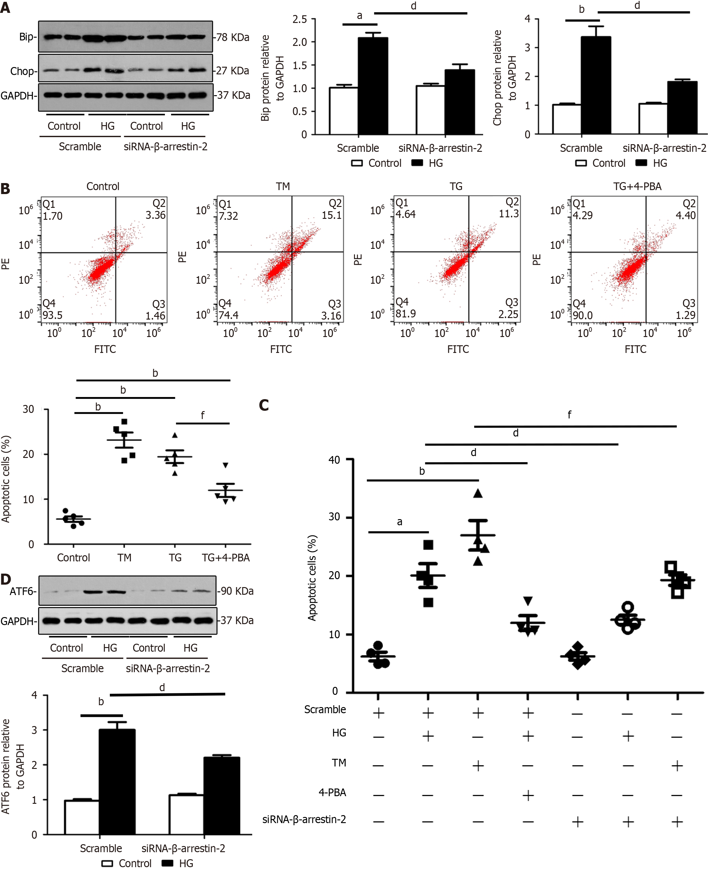
Figure 4 Knockdown of β-arrestin-2 reduces endoplasmic reticulum stress and apoptosis of glomerular endothelial cells in high glucose treatments by decreasing expression of activating transcription factor 6.
A: Representative images and summarized data showing that knockdown of β-arrestin-2 inactivated endoplasmic reticulum (ER) stress by downregulating expression of BiP and CHOP in HG-treated glomerular endothelial cells (GENCs) by Western blotting (aP < 0.05 vs scramble/control, bP < 0.01 vs scramble/control, dP < 0.05 vs scramble/HG treatment, n = 6); B: Flow cytometric analysis showing that the ER stress activators tunicamycin (TM) and thapsigargin (TG) induced GENC apoptosis and the inhibitor 4-phenyl butyric acid (4-PBA) reduced GENC apoptosis (bP < 0.01 vs scramble/control, fP < 0.05 vs TM/TG treatment, n = 5); C: Summarized flow cytometric data showing apoptosis of GENCs under different stimuli (aP < 0.05 vs scramble/control, bP < 0.01 vs scramble/control, dP < 0.05 vs scramble/HG treatment, fP < 0.05 vs TM/TG treatment, n = 4); D: Western blotting images showing effects of β-arrestin-2 knockdown on expression of ATF6 in GENCs under HG treatment (bP < 0.01 vs scramble/control, dP < 0.05 vs scramble/HG treatment, n = 6). HG: High glucose; siRNA: Small interfering RNA; TM: Tunicamycin; TG: Thapsigargin; 4-PBA: 4-Phenyl butyric acid.
- Citation: Liu J, Song XY, Li XT, Yang M, Wang F, Han Y, Jiang Y, Lei YX, Jiang M, Zhang W, Tang DQ. β-Arrestin-2 enhances endoplasmic reticulum stress-induced glomerular endothelial cell injury by activating transcription factor 6 in diabetic nephropathy. World J Diabetes 2024; 15(12): 2322-2337
- URL: https://www.wjgnet.com/1948-9358/full/v15/i12/2322.htm
- DOI: https://dx.doi.org/10.4239/wjd.v15.i12.2322


