Copyright
©The Author(s) 2024.
World J Diabetes. Dec 15, 2024; 15(12): 2322-2337
Published online Dec 15, 2024. doi: 10.4239/wjd.v15.i12.2322
Published online Dec 15, 2024. doi: 10.4239/wjd.v15.i12.2322
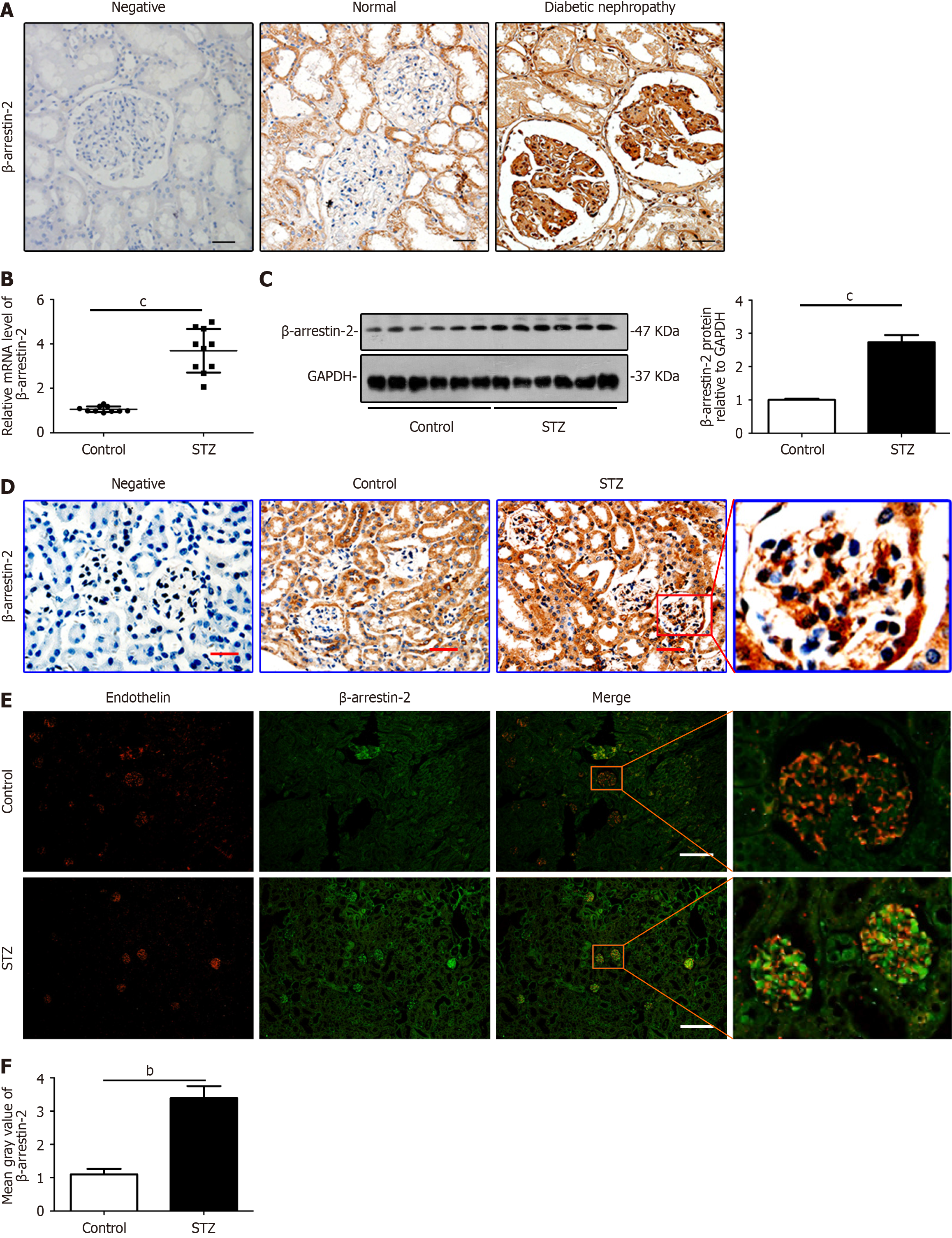
Figure 1 β-Arrestin-2 is increased significantly in renal biopsies from patients with diabetic nephropathy and glomerular endothelial cells from mice with diabetic nephropathy.
A: Images of immunohistochemical (IHC) staining to detect the expression of β-arrestin-2 in paraffin sections of human renal tissue from normal controls and patients with diabetic nephropathy (DN) (black bars = 10 μm, n = 5); B: Relative mRNA levels of β-arrestin-2 in the renal cortex of DN mice (mean ± SD, cP < 0.001 vs control, n = 8); C: The expression of β-arrestin-2 in the renal cortex of DN mice was analyzed by immunoblotting (cP < 0.001 vs control, n = 8); D: Representative images of IHC staining to detect the expression of β-arrestin-2 in paraffin sections of the kidneys from control and DN mice (red bars = 20 μm, n = 8); E: Detection of β-arrestin-2 expression in glomerular endothelial cells (GENCs) in streptozotocin (STZ)-induced DN mouse model by immunofluorescence double labeling: Endothelin (red, mark protein in GENCs) and β-arrestin-2 (green) (white bars = 50 μm, n = 8); F: Quantifications showing expression of β-arrestin-2 in the kidneys from different groups of mice (bP < 0.01 vs control, n = 8).
- Citation: Liu J, Song XY, Li XT, Yang M, Wang F, Han Y, Jiang Y, Lei YX, Jiang M, Zhang W, Tang DQ. β-Arrestin-2 enhances endoplasmic reticulum stress-induced glomerular endothelial cell injury by activating transcription factor 6 in diabetic nephropathy. World J Diabetes 2024; 15(12): 2322-2337
- URL: https://www.wjgnet.com/1948-9358/full/v15/i12/2322.htm
- DOI: https://dx.doi.org/10.4239/wjd.v15.i12.2322


