Copyright
©The Author(s) 2024.
World J Diabetes. Jan 15, 2024; 15(1): 92-104
Published online Jan 15, 2024. doi: 10.4239/wjd.v15.i1.92
Published online Jan 15, 2024. doi: 10.4239/wjd.v15.i1.92
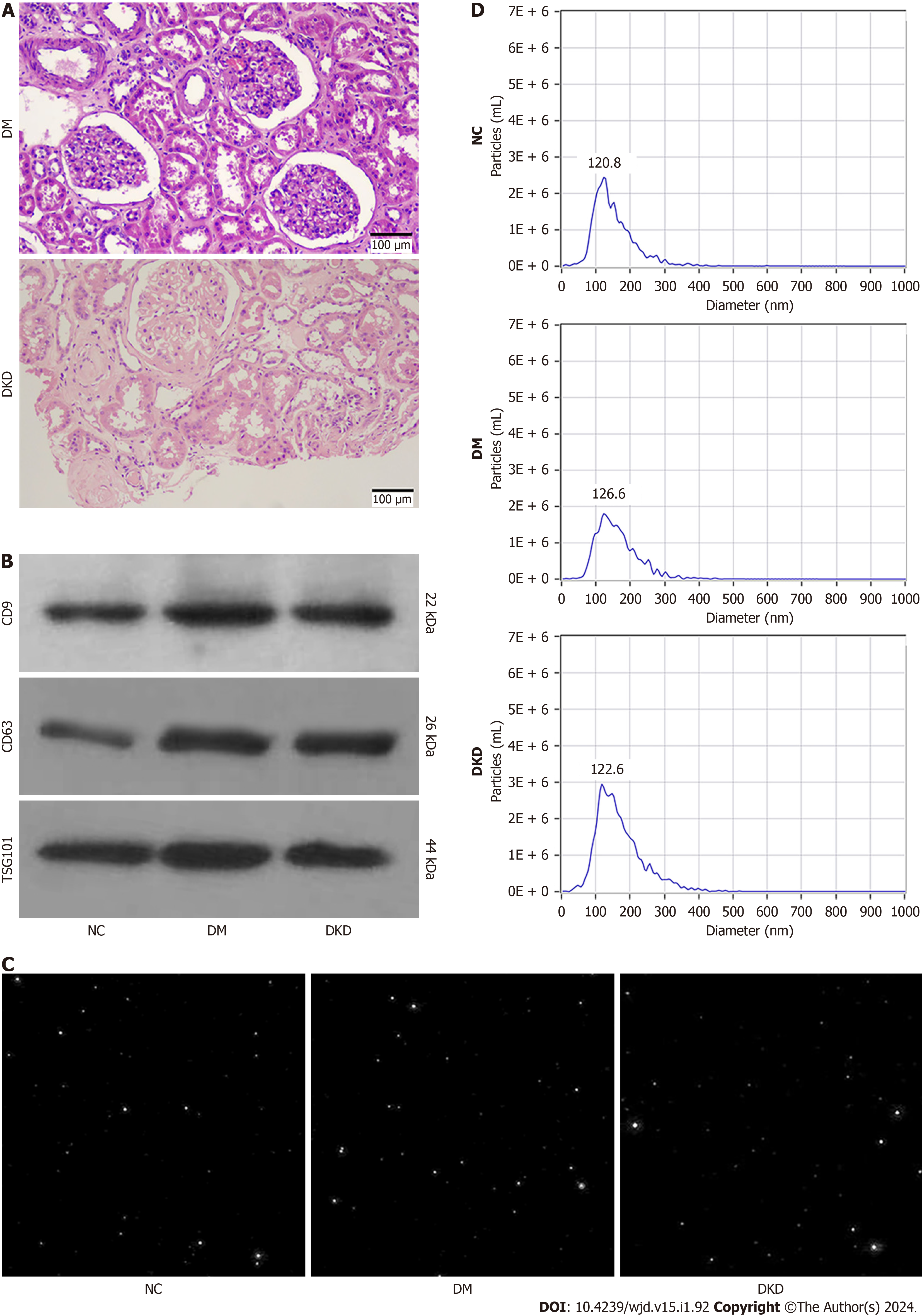
Figure 1 The characterization of urinary exosome.
A: The glomeruli histological features of diabetic kidney disease patients (Periodic acid-Schiff staining), Bar = 100 μm; B: The exosomal surface markers CD9, CD63 and TSG101 were detected by western blotting; C: The particle screenshots of nanoparticle tracking analysis (NTA) in the urine samples from participants; D: The diameter sizes and diameter concentration distributions of the particles were measured by NTA. NC: Normal control; DM: Diabetes mellitus; DKD: Diabetic kidney disease.
- Citation: Han LL, Wang SH, Yao MY, Zhou H. Urinary exosomal microRNA-145-5p and microRNA-27a-3p act as noninvasive diagnostic biomarkers for diabetic kidney disease. World J Diabetes 2024; 15(1): 92-104
- URL: https://www.wjgnet.com/1948-9358/full/v15/i1/92.htm
- DOI: https://dx.doi.org/10.4239/wjd.v15.i1.92


