Copyright
©The Author(s) 2024.
World J Diabetes. Jan 15, 2024; 15(1): 105-125
Published online Jan 15, 2024. doi: 10.4239/wjd.v15.i1.105
Published online Jan 15, 2024. doi: 10.4239/wjd.v15.i1.105
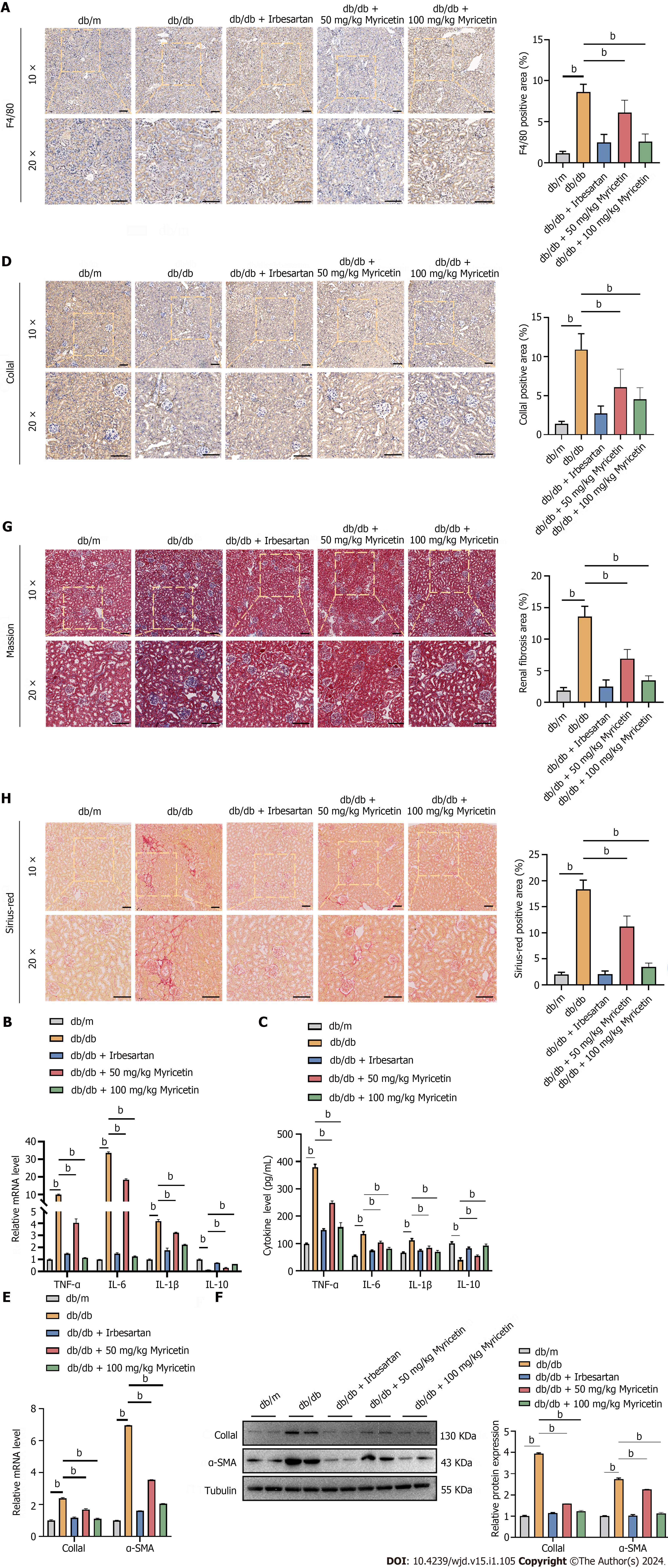
Figure 3 Myricetin inhibited inflammation factors and fibrosis in the renal tissue of diabetic nephropathy mice.
A: Immunofluorescent staining of F4/80 protein; B: Tumor necrosis factor-alpha (TNF-α), interleukin (IL)-6, IL-1β, and IL-10 mRNA levels; C: Serum TNF-α, IL-6, IL-1β, and IL-10 Levels; D: Immunofluorescent staining of collagen-1a1 (Col1a1) protein; E: Reverse transcription-PCR analysis of Col1a1 and alpha-smooth muscle actin (α-SMA) mRNAs; F: Western blotting analysis of Col1a1 and α-SMA proteins; G: Masson’s trichrome staining (left) and renal fibrosis analysis (right); H: Sirius-red staining (left) and fibrosis analysis (right). TNF-α: Tumor necrosis factor-alpha; α-SMA: Alpha-smooth muscle actin; IL: Interleukin; Col1a1: Collagen-1a1. bP < 0.05.
- Citation: Xu WL, Zhou PP, Yu X, Tian T, Bao JJ, Ni CR, Zha M, Wu X, Yu JY. Myricetin induces M2 macrophage polarization to alleviate renal tubulointerstitial fibrosis in diabetic nephropathy via PI3K/Akt pathway. World J Diabetes 2024; 15(1): 105-125
- URL: https://www.wjgnet.com/1948-9358/full/v15/i1/105.htm
- DOI: https://dx.doi.org/10.4239/wjd.v15.i1.105


