Copyright
©The Author(s) 2023.
World J Diabetes. Jul 15, 2023; 14(7): 1013-1026
Published online Jul 15, 2023. doi: 10.4239/wjd.v14.i7.1013
Published online Jul 15, 2023. doi: 10.4239/wjd.v14.i7.1013
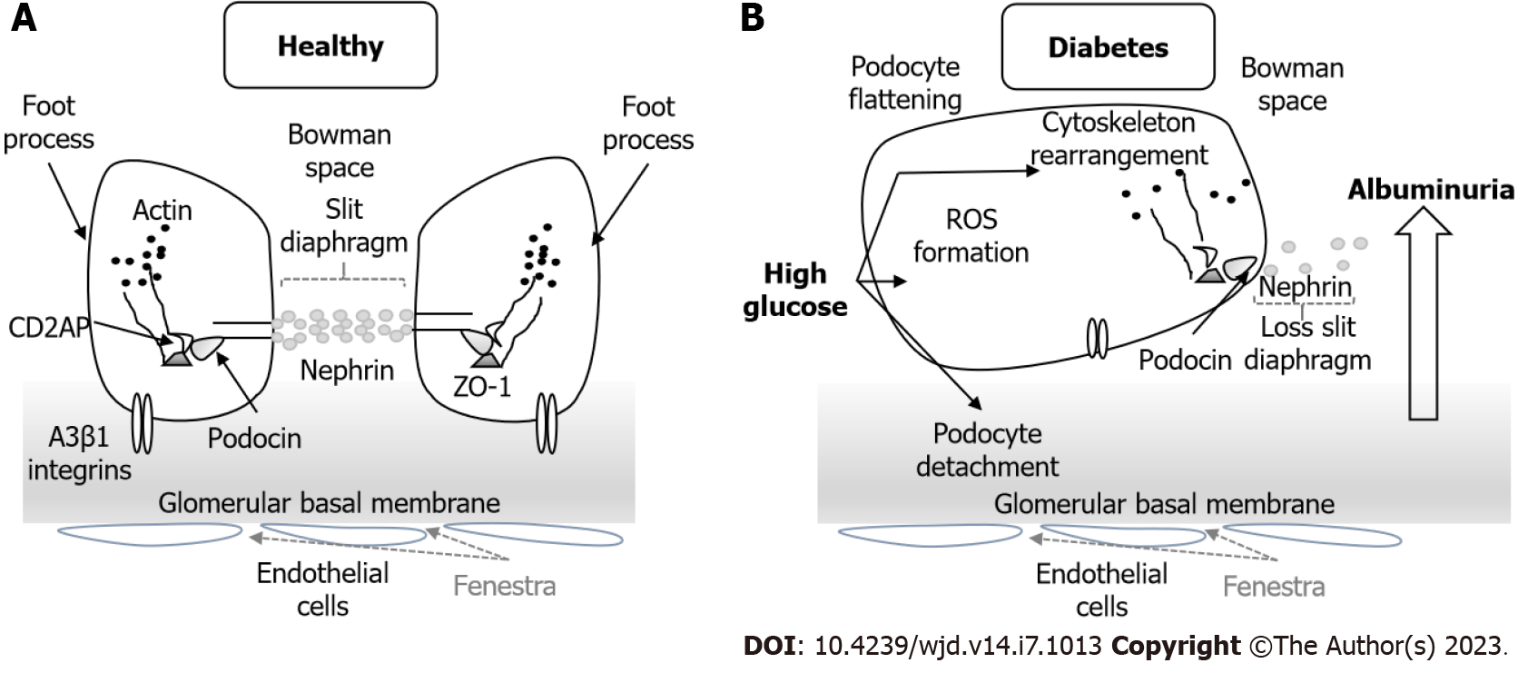
Figure 6 Podocyte structure.
A: Normal structure of the podocyte with the morphology of the foot process and the slit diaphragm; B: In diabetic nephropathy the podocyte structure and slit diaphragm are injured with slit diaphragm disruption, podocyte detachment and cytoskeleton rearrangement. These changes lead to albuminuria and progressive kidney disease. ROS: Reactive oxygen species; ZO: Zonula occludens.
- Citation: Robles-Osorio ML, Sabath E. Tight junction disruption and the pathogenesis of the chronic complications of diabetes mellitus: A narrative review. World J Diabetes 2023; 14(7): 1013-1026
- URL: https://www.wjgnet.com/1948-9358/full/v14/i7/1013.htm
- DOI: https://dx.doi.org/10.4239/wjd.v14.i7.1013


