Copyright
©The Author(s) 2023.
World J Diabetes. Jan 15, 2023; 14(1): 48-61
Published online Jan 15, 2023. doi: 10.4239/wjd.v14.i1.48
Published online Jan 15, 2023. doi: 10.4239/wjd.v14.i1.48
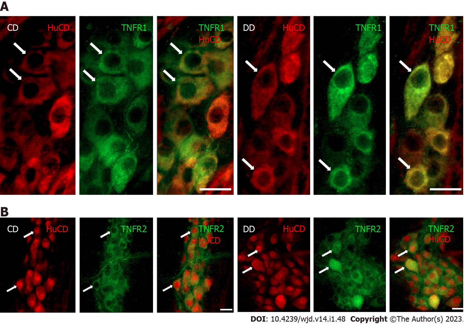
Figure 1 Representative fluorescent micrographs of whole-mount preparations of myenteric ganglia from the duodenum of a control and a diabetic rat after TNFR1-HuCD or TNFR2-HuCD double-labeling immunohistochemistry.
HuCD as a pan-neuronal marker was applied to label myenteric neurons. A: TNFR1-HuCD; B: TNFR2-HuCD. Arrows indicate myenteric neurons. Scale bar: 20 μm. CD: Control duodenum; DD: Diabetic duodenum.
- Citation: Barta BP, Onhausz B, AL Doghmi A, Szalai Z, Balázs J, Bagyánszki M, Bódi N. Gut region-specific TNFR expression: TNFR2 is more affected than TNFR1 in duodenal myenteric ganglia of diabetic rats. World J Diabetes 2023; 14(1): 48-61
- URL: https://www.wjgnet.com/1948-9358/full/v14/i1/48.htm
- DOI: https://dx.doi.org/10.4239/wjd.v14.i1.48


