Copyright
©The Author(s) 2022.
World J Diabetes. Sep 15, 2022; 13(9): 765-775
Published online Sep 15, 2022. doi: 10.4239/wjd.v13.i9.765
Published online Sep 15, 2022. doi: 10.4239/wjd.v13.i9.765
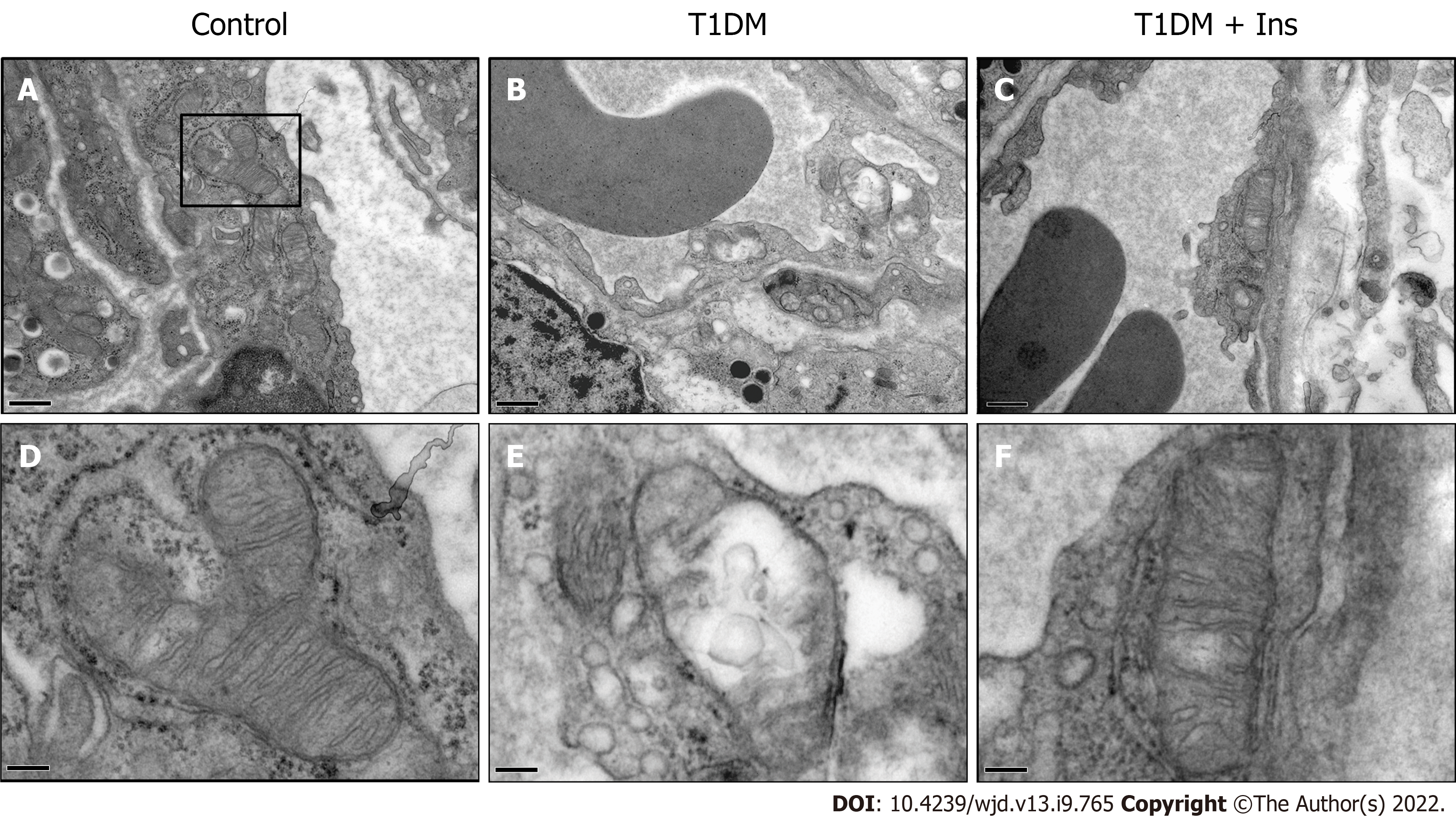
Figure 2 Glucotoxicity induced ultrastructural damage to mitochondria in islet microvascular endothelial cells.
The ultrastructure of pancreatic islet microvascular endothelial cells (IMECs) in the control (A), type 1 diabetes mellitus (T1DM) (B) and insulin-treated groups (C) was revealed by TEM (upper panels, scale bar = 0.5 μm). The ultrastructure of mitochondria in IMECs in the control (D), T1DM (E) and insulin-treated groups (F) is shown in the lower panels. Swollen mitochondria with cristae rupture or disappearance and a transparent matrix were found in T1DM mice. Restored mitochondria were observed after insulin administration (lower panels, scale bar = 2 μm).
- Citation: Li BW, Li Y, Zhang X, Fu SJ, Wang B, Zhang XY, Liu XT, Wang Q, Li AL, Liu MM. Role of insulin in pancreatic microcirculatory oxygen profile and bioenergetics. World J Diabetes 2022; 13(9): 765-775
- URL: https://www.wjgnet.com/1948-9358/full/v13/i9/765.htm
- DOI: https://dx.doi.org/10.4239/wjd.v13.i9.765


