Copyright
©The Author(s) 2022.
World J Diabetes. Dec 15, 2022; 13(12): 1131-1139
Published online Dec 15, 2022. doi: 10.4239/wjd.v13.i12.1131
Published online Dec 15, 2022. doi: 10.4239/wjd.v13.i12.1131
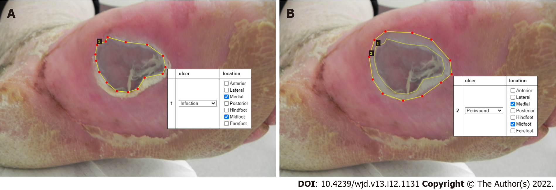
Figure 2 An infected ulcer on the plantar aspect of the left foot.
A: The ulcer is labelled (dotted line marking the boundaries) for the training set. The white box shows the site and type of ulcer. B: The peri-wound of the ulcer is labelled with a dotted line. The white box shows the site of the ulcer and the peri-wound.
- Citation: Pappachan JM, Cassidy B, Fernandez CJ, Chandrabalan V, Yap MH. The role of artificial intelligence technology in the care of diabetic foot ulcers: the past, the present, and the future. World J Diabetes 2022; 13(12): 1131-1139
- URL: https://www.wjgnet.com/1948-9358/full/v13/i12/1131.htm
- DOI: https://dx.doi.org/10.4239/wjd.v13.i12.1131


