Copyright
©The Author(s) 2021.
World J Diabetes. Jun 15, 2021; 12(6): 745-766
Published online Jun 15, 2021. doi: 10.4239/wjd.v12.i6.745
Published online Jun 15, 2021. doi: 10.4239/wjd.v12.i6.745
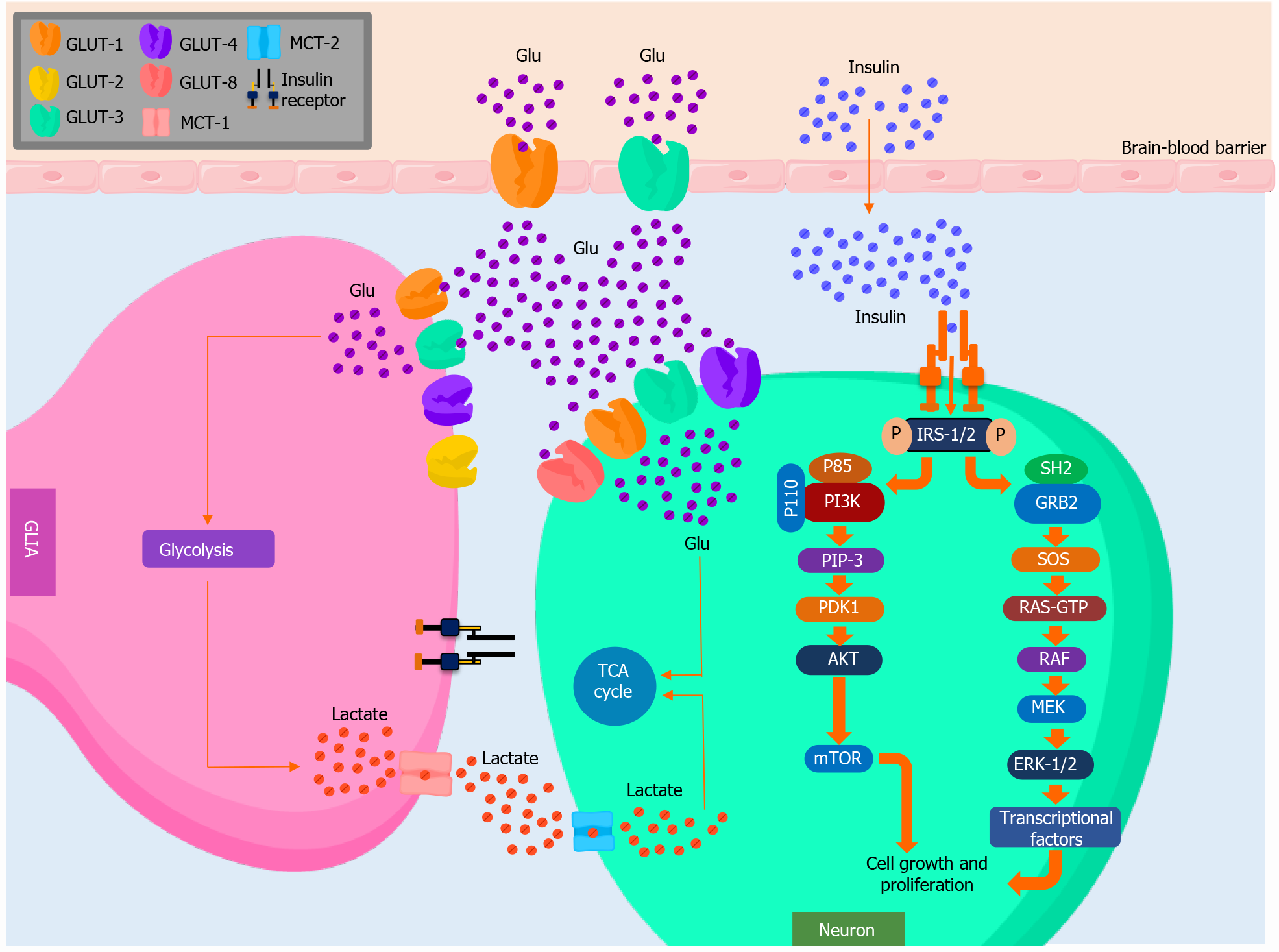
Figure 1 Molecular basis of brain glucose and insulin metabolism.
Glucose transporter (GLUT)-1 and GLUT-3 are widely expressed in the brain blood barrier (BBB), allowing glucose transport. Likewise, GLUT-1, GLUT-2, GLUT-3, and GLUT-4 are present in the membrane of glial cells, whereas GLUT-1, GLUT-3, GLUT-4, and GLUT-8 are expressed in neurons. These allow the transport of glucose to the intracellular environment, ready for energetic metabolism. On the other hand, it has been proposed that insulin crosses the BBB through a saturable transporter and then couples to the insulin receptor substrate (IRS) in the neuronal membrane, causing a conformational change that phosphorylates IRS-1/2, which mainly activates the AKT and extracellular signal-regulated kinase (ERK)-1/2 pathways. After a phosphorylation cascade, this activates mTOR and various transcriptional factors involved in the growth and cellular differentiation of the nervous system. Glu: Glucose; GLUT: Glucose transporters; IR: Insulin receptor; MCT: Monocarboxylate transporter; IRS: Insulin receptor substrate; GRB2: Growth factor receptor-bound protein 2; SOS: Son of Sevenless homolog; RAF: Rapidly accelerated fibrosarcoma kinase; MEK: Mitogen-activated protein kinases; ERK: Extracellular signal-regulated kinase; PI3K: Phosphatidylinositol 3-kinase; PIP-3: Phosphatidylinositol (3,4,5)-trisphosphate; PDK1: Phosphoinositide-dependent kinase-1; AKT: Protein kinase B; TCA cycle: Tricarboxylic acid cycle.
- Citation: Rojas M, Chávez-Castillo M, Bautista J, Ortega Á, Nava M, Salazar J, Díaz-Camargo E, Medina O, Rojas-Quintero J, Bermúdez V. Alzheimer’s disease and type 2 diabetes mellitus: Pathophysiologic and pharmacotherapeutics links. World J Diabetes 2021; 12(6): 745-766
- URL: https://www.wjgnet.com/1948-9358/full/v12/i6/745.htm
- DOI: https://dx.doi.org/10.4239/wjd.v12.i6.745


