Copyright
©The Author(s) 2021.
World J Diabetes. Apr 15, 2021; 12(4): 437-452
Published online Apr 15, 2021. doi: 10.4239/wjd.v12.i4.437
Published online Apr 15, 2021. doi: 10.4239/wjd.v12.i4.437
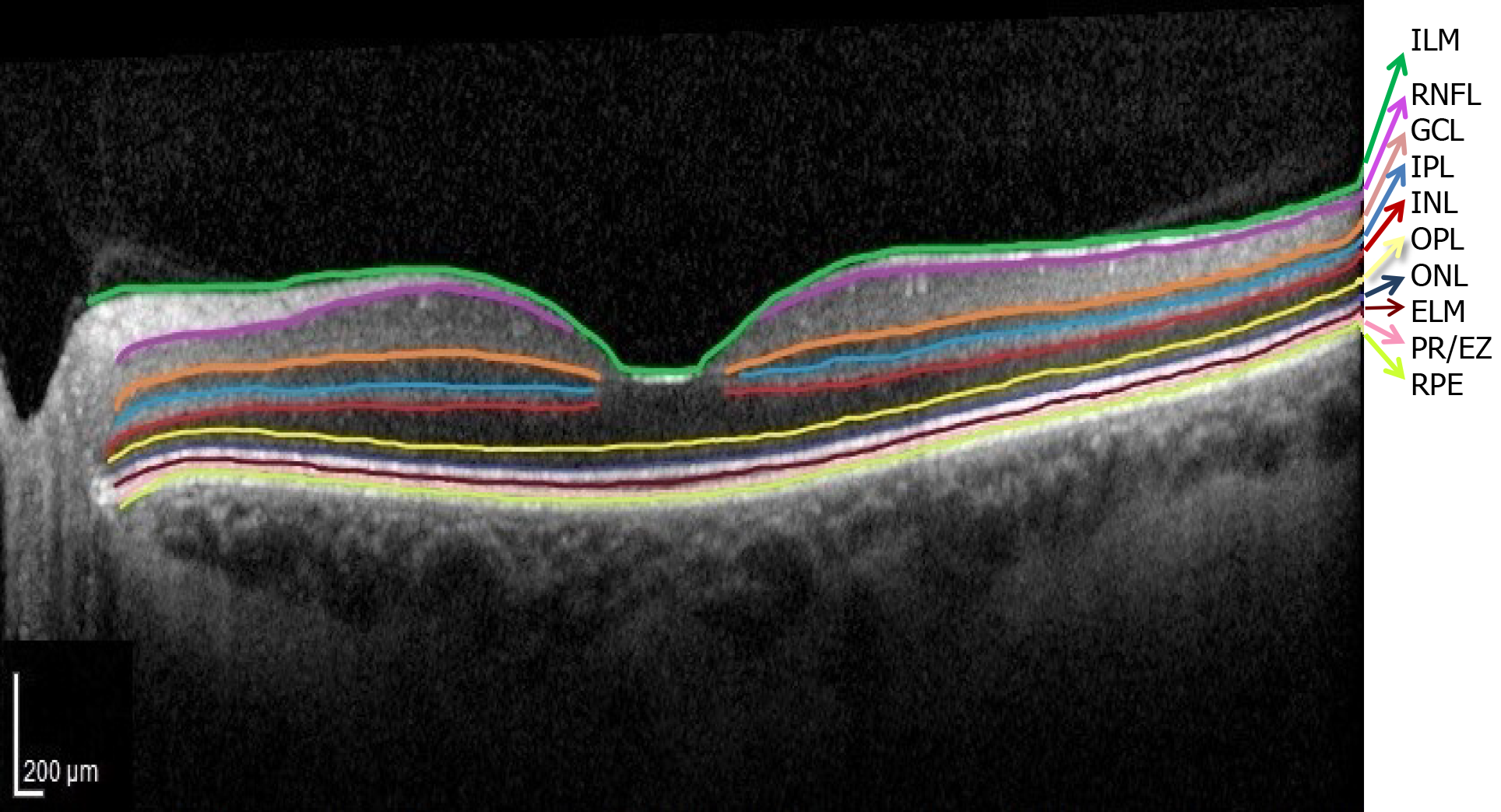
Figure 3 Normal optical coherence tomography aspect of the retinal layers.
Segmentation software automatically marked the 10 retinal layers. (ILM: Internal limiting membrane; RNFL: Retinal nerve fiber layer; GCL: Ganglion cell layer; IPL: Inner plexiform layer; INL: Inner nuclear layer; OPL: Outer plexiform layer; ONL: Outer nuclear layer; ELM: External limiting membrane; PR/EZ: Photoreceptor layer/ellipsoid zone (inner and outer photoreceptor segment junction; RPE: Retinal pigment epithelium).
- Citation: Ţălu Ş, Nicoara SD. Malfunction of outer retinal barrier and choroid in the occurrence and progression of diabetic macular edema . World J Diabetes 2021; 12(4): 437-452
- URL: https://www.wjgnet.com/1948-9358/full/v12/i4/437.htm
- DOI: https://dx.doi.org/10.4239/wjd.v12.i4.437


