Copyright
©2014 Baishideng Publishing Group Co.
World J Diabetes. Apr 15, 2014; 5(2): 219-223
Published online Apr 15, 2014. doi: 10.4239/wjd.v5.i2.219
Published online Apr 15, 2014. doi: 10.4239/wjd.v5.i2.219
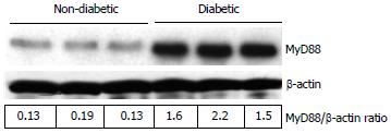
Figure 1 Representative Western blot showing the MyD88 protein expression in non-diabetic and diabetic wound tissues.
Wound tissues were collected, lysed and 25 μg protein was blotted for MyD88 and β-actin. Densitometric ratios (MyD88/β-actin) are indicated below. Each lane presents protein from an individual patient wound debridement tissue (n = 3/group).
Figure 2 The DNA-binding activity of nuclear nuclear factor-kappa B p65 in wound tissues was determined using ELISA technique.
Values are normalized to mg nuclear protein and expressed as mean ± SD. bP < 0.001 vs non-diabetic.
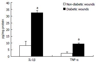
Figure 3 Interleukin-1β and tumor necrosis factor-α concentration in wound tissues were determined by ELISA assay.
Values are normalized to mg protein and expressed as mean ± SD. aP < 0.05 vs non-diabetic. IL-1β: Interleukin-1beta; TNF-α: Tumor necrosis factor-alpha.
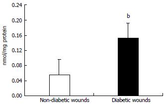
Figure 4 Lipid peroxidation in wound tissue lysates were determined using thiobarbituric acid reactive substances assay as described in Materials and methods.
Values are normalized to mg protein and expressed as mean ± SD. bP < 0.01 vs non-diabetic.
- Citation: Dasu MR, Martin SJ. Toll-like receptor expression and signaling in human diabetic wounds. World J Diabetes 2014; 5(2): 219-223
- URL: https://www.wjgnet.com/1948-9358/full/v5/i2/219.htm
- DOI: https://dx.doi.org/10.4239/wjd.v5.i2.219


