Copyright
©The Author(s) 2017.
World J Gastrointest Oncol. Sep 15, 2017; 9(9): 390-396
Published online Sep 15, 2017. doi: 10.4251/wjgo.v9.i9.390
Published online Sep 15, 2017. doi: 10.4251/wjgo.v9.i9.390
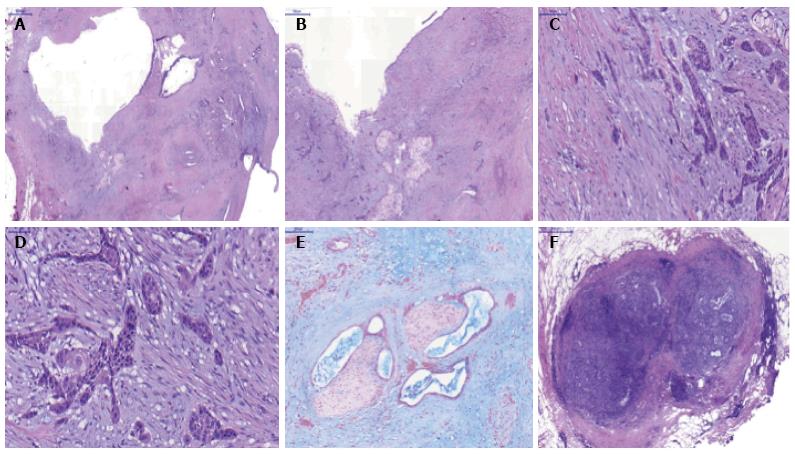
Figure 4 Microscopic pathology of the surgical specimen.
A: Intraductal papillary mucinous neoplasm with adenocarcinoma component, hematoxylin and eosin (H and E) 10 ×; B-D: Squamous metaplasia and evident infiltrative squamous carcinoma, H and E 10 ×, 20 × and 40 ×, respectively; E: Adenocarcinoma with perineural invasion, alcian blue 20 ×; F: Peripancreatic lymph node metastasis (adenocarcinoma component), H and E 20 ×.
- Citation: Martínez de Juan F, Reolid Escribano M, Martínez Lapiedra C, Maia de Alcantara F, Caballero Soto M, Calatrava Fons A, Machado I. Pancreatic adenosquamous carcinoma and intraductal papillary mucinous neoplasm in a CDKN2A germline mutation carrier. World J Gastrointest Oncol 2017; 9(9): 390-396
- URL: https://www.wjgnet.com/1948-5204/full/v9/i9/390.htm
- DOI: https://dx.doi.org/10.4251/wjgo.v9.i9.390


