Copyright
©The Author(s) 2017.
World J Gastrointest Oncol. Sep 15, 2017; 9(9): 390-396
Published online Sep 15, 2017. doi: 10.4251/wjgo.v9.i9.390
Published online Sep 15, 2017. doi: 10.4251/wjgo.v9.i9.390
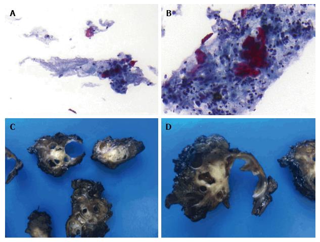
Figure 3 Endoscopic ultrasonography fine needle aspiration biopsies and surgical specimen.
A and B: Positive cytology from the pancreatic mass (adenocarcinoma with a significant keratinizing component suggestive of adenosquamous carcinoma), Papanicolaou staining 20 × and 40 ×, respectively; C and D: A solid-cystic pancreatic mass (gross pathology).
- Citation: Martínez de Juan F, Reolid Escribano M, Martínez Lapiedra C, Maia de Alcantara F, Caballero Soto M, Calatrava Fons A, Machado I. Pancreatic adenosquamous carcinoma and intraductal papillary mucinous neoplasm in a CDKN2A germline mutation carrier. World J Gastrointest Oncol 2017; 9(9): 390-396
- URL: https://www.wjgnet.com/1948-5204/full/v9/i9/390.htm
- DOI: https://dx.doi.org/10.4251/wjgo.v9.i9.390


