Copyright
©The Author(s) 2017.
World J Gastrointest Oncol. Nov 15, 2017; 9(11): 444-451
Published online Nov 15, 2017. doi: 10.4251/wjgo.v9.i11.444
Published online Nov 15, 2017. doi: 10.4251/wjgo.v9.i11.444
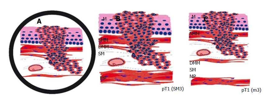
Figure 1 The difficulty in distinguishing between the smooth muscle cells of the muscularis mucosae, which can eventually be splintered in the superficial muscularis mucosae and the deep muscularis mucosae, and those of the muscularis propria.
This schematic drawing shows a section of a Barrett’s adenocarcinoma that may, at first glance appear, relatively straightforward, but determining the type of muscle layer can be challenging. Identifying large vessels (A) may suggest the diagnosis of submucosal invasion (B) and is, therefore, a well-known pitfall, as large vessels can also be found in between the superficial and DMM (C). An intramucosal carcinoma pT1 (m3) can therefore easily be mistaken as a submucosal carcinoma pT1 (sm). M: Mucosa; SMM: Superficial muscularis mucosae; DMM: Deep muscularis mucosae; MP: Muscularis propria.
- Citation: Endhardt K, Märkl B, Probst A, Schaller T, Aust D. Value of histomorphometric tumour thickness and smoothelin for conventional m-classification in early oesophageal adenocarcinoma. World J Gastrointest Oncol 2017; 9(11): 444-451
- URL: https://www.wjgnet.com/1948-5204/full/v9/i11/444.htm
- DOI: https://dx.doi.org/10.4251/wjgo.v9.i11.444


