Copyright
©The Author(s) 2016.
World J Gastrointest Oncol. Aug 15, 2016; 8(8): 615-622
Published online Aug 15, 2016. doi: 10.4251/wjgo.v8.i8.615
Published online Aug 15, 2016. doi: 10.4251/wjgo.v8.i8.615
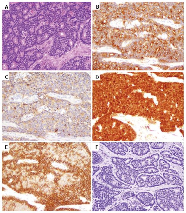
Figure 2 A pancreatic neuroendocrine neoplasm from a familial adenomatous polyposis patient (original magnification 200 ×).
A: Hematoxylin and eosin stain showing a well differentiated neuroendocrine tumor composed of monomorphic cells with “salt and pepper” chromatin; B and C: Immunohistochemical labeling for synaptophysin (B) and chromogranin (C) showing positive staining; D: Immunohistochemical labeling with β catenin showing β catenin nuclear accumulation and loss of membrane staining; E: Immunohistochemical staining for E-cadherin showing strong E-cadherin membranous expression; F: Immunohistochemical labeling for APC showing loss of APC expression. APC: Adenomatous polyposis coli.
- Citation: Weiss V, Dueber J, Wright JP, Cates J, Revetta F, Parikh AA, Merchant NB, Shi C. Immunohistochemical analysis of the Wnt/β-catenin signaling pathway in pancreatic neuroendocrine neoplasms. World J Gastrointest Oncol 2016; 8(8): 615-622
- URL: https://www.wjgnet.com/1948-5204/full/v8/i8/615.htm
- DOI: https://dx.doi.org/10.4251/wjgo.v8.i8.615


