Copyright
©The Author(s) 2016.
World J Gastrointest Oncol. Aug 15, 2016; 8(8): 615-622
Published online Aug 15, 2016. doi: 10.4251/wjgo.v8.i8.615
Published online Aug 15, 2016. doi: 10.4251/wjgo.v8.i8.615
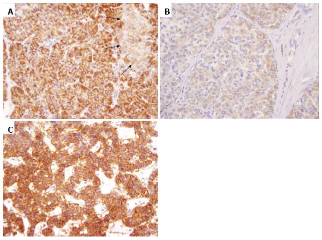
Figure 1 β-catenin expression in the pancreas and pancreatic neuroendocrine neoplasms (original magnification 200 ×).
A: Strong membranous β-catenin staining in the exocrine pancreas and weak membranous staining in pancreatic islets (black arrows); B: Weak membranous β-catenin staining in one pancreatic neuroendocrine neoplasm; C: Strong membranous β-catenin staining in another pancreatic neuroendocrine neoplasm.
- Citation: Weiss V, Dueber J, Wright JP, Cates J, Revetta F, Parikh AA, Merchant NB, Shi C. Immunohistochemical analysis of the Wnt/β-catenin signaling pathway in pancreatic neuroendocrine neoplasms. World J Gastrointest Oncol 2016; 8(8): 615-622
- URL: https://www.wjgnet.com/1948-5204/full/v8/i8/615.htm
- DOI: https://dx.doi.org/10.4251/wjgo.v8.i8.615


