Copyright
©The Author(s) 2016.
World J Gastrointest Oncol. Mar 15, 2016; 8(3): 271-281
Published online Mar 15, 2016. doi: 10.4251/wjgo.v8.i3.271
Published online Mar 15, 2016. doi: 10.4251/wjgo.v8.i3.271
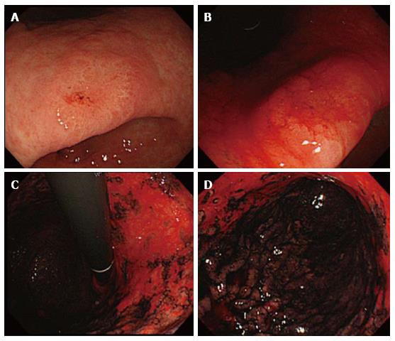
Figure 1 Representative photos of Congo-red chromoendoscopy in a 66-year-old female patient with gastric cancer that emerged after successful eradication of Helicobacter pylori infection.
A: A lesion of well-differentiated adenocarcinoma located on the lesser curvature in the lower part of the stomach [FQ260Z (Olympus, Tokyo)]; B: Congo-red imaging showed GC in a non-acid secreting area (Red); C and D: Post-eradicated gastric background mucosa (Red: Non-acid secreting area; Black: Acid secreting area). GC: Gastric cancer.
- Citation: Uno K, Iijima K, Shimosegawa T. Gastric cancer development after the successful eradication of Helicobacter pylori. World J Gastrointest Oncol 2016; 8(3): 271-281
- URL: https://www.wjgnet.com/1948-5204/full/v8/i3/271.htm
- DOI: https://dx.doi.org/10.4251/wjgo.v8.i3.271


