Copyright
©The Author(s) 2015.
World J Gastrointest Oncol. Aug 15, 2015; 7(8): 118-122
Published online Aug 15, 2015. doi: 10.4251/wjgo.v7.i8.118
Published online Aug 15, 2015. doi: 10.4251/wjgo.v7.i8.118
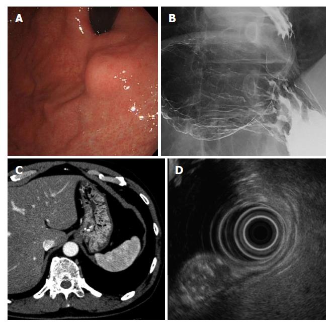
Figure 1 Results of pre-operative examinations.
A: Endoscopic findings showing a submucosal lesion of 15 mm in diameter on the anterior wall of the upper gastric body near the esophago-gastric junction. The surface was covered with normal gastric mucosa; B: Barium gastrography showed a smooth elevated lesion of 2 cm in diameter on the anterior wall of the upper gastric body near the esophago-gastric junction; C: Computed tomography revealed a 15-mm submucosal low density area with calcification in the anterior wall of the upper gastric body. No lymph node or distant metastasis was detected; D: Endoscopic ultrasound showed an 11.2 mm × 13.5 mm submucosal tumor derived from the third layer of the gastric wall as a heterogeneous lesion with a mixture of a high echoic lesion, low echoic lesion, and calcification.
- Citation: Imamura T, Komatsu S, Ichikawa D, Kobayashi H, Miyamae M, Hirajima S, Kawaguchi T, Kubota T, Kosuga T, Okamoto K, Konishi H, Shiozaki A, Fujiwara H, Ogiso K, Yagi N, Yanagisawa A, Ando T, Otsuji E. Gastric carcinoma originating from the heterotopic submucosal gastric gland treated by laparoscopy and endoscopy cooperative surgery. World J Gastrointest Oncol 2015; 7(8): 118-122
- URL: https://www.wjgnet.com/1948-5204/full/v7/i8/118.htm
- DOI: https://dx.doi.org/10.4251/wjgo.v7.i8.118


