Copyright
©2014 Baishideng Publishing Group Inc.
World J Gastrointest Oncol. Sep 15, 2014; 6(9): 351-359
Published online Sep 15, 2014. doi: 10.4251/wjgo.v6.i9.351
Published online Sep 15, 2014. doi: 10.4251/wjgo.v6.i9.351
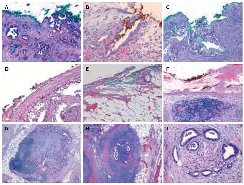
Figure 3 Microscopic picture.
A-C: Microscopic picture of tumor glands in direct contact with an inked margin (R1 resection) (HE × 200, × 400 and × 200, respectively); D: Neoplastic cells within 1 mm of the resection margin colored in black (HE × 200); E, F: Examples of free medial or posterior margin (HE × 200); G: Ganglionar metastases (HE × 200); H: Vascular invasion (HE × 200); I: Perineural invasion (HE × 400).
- Citation: Gómez-Mateo MDC, Sabater-Ortí L, Ferrández-Izquierdo A. Pathology handling of pancreatoduodenectomy specimens: Approaches and controversies. World J Gastrointest Oncol 2014; 6(9): 351-359
- URL: https://www.wjgnet.com/1948-5204/full/v6/i9/351.htm
- DOI: https://dx.doi.org/10.4251/wjgo.v6.i9.351


