Copyright
©2014 Baishideng Publishing Group Inc.
World J Gastrointest Oncol. Sep 15, 2014; 6(9): 330-343
Published online Sep 15, 2014. doi: 10.4251/wjgo.v6.i9.330
Published online Sep 15, 2014. doi: 10.4251/wjgo.v6.i9.330
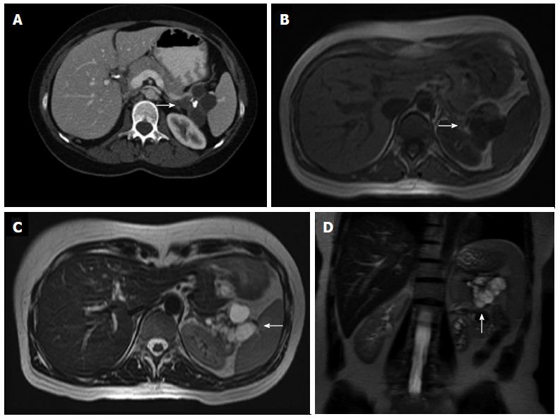
Figure 18 Axial nonenhanced multidetector computed tomography image.
A: A polylobulated cystic lesion with a coarse calcification in its center (arrow), which is the phathognomonic central scar for serous cystadenoma; B-D: Magnetic resonance imaging show a cluster of small cysts (arrows), which are hypointense in T1-weighted images (B) and hyperintense in T2-weighted images (C, D), without visible communication within the cyst or the pancreatic duct. A central signal void is also identifiable.
- Citation: Santa LGL, Retortillo JAP, Miguel AC, Klein LM. Radiology of pancreatic neoplasms: An update. World J Gastrointest Oncol 2014; 6(9): 330-343
- URL: https://www.wjgnet.com/1948-5204/full/v6/i9/330.htm
- DOI: https://dx.doi.org/10.4251/wjgo.v6.i9.330


