Copyright
©2014 Baishideng Publishing Group Inc.
World J Gastrointest Oncol. Sep 15, 2014; 6(9): 330-343
Published online Sep 15, 2014. doi: 10.4251/wjgo.v6.i9.330
Published online Sep 15, 2014. doi: 10.4251/wjgo.v6.i9.330
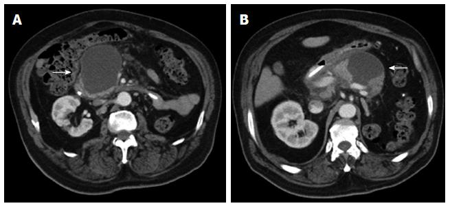
Figure 17 Axial contrast enhanced multidetector computed tomography images (A, B) reveal a homogeneously hypodense intraparenchymal fluid collection of the pancreas without any non-liquefied material in it, encapsulated completely by a thin slightly hyperdense layer (arrows).
These findings are compatible with a pseudocyst in a patient with a clinical history of pancreatitis.
- Citation: Santa LGL, Retortillo JAP, Miguel AC, Klein LM. Radiology of pancreatic neoplasms: An update. World J Gastrointest Oncol 2014; 6(9): 330-343
- URL: https://www.wjgnet.com/1948-5204/full/v6/i9/330.htm
- DOI: https://dx.doi.org/10.4251/wjgo.v6.i9.330


