Copyright
©2014 Baishideng Publishing Group Inc.
World J Gastrointest Oncol. Sep 15, 2014; 6(9): 330-343
Published online Sep 15, 2014. doi: 10.4251/wjgo.v6.i9.330
Published online Sep 15, 2014. doi: 10.4251/wjgo.v6.i9.330
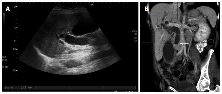
Figure 7 Indirect signs of pancreatic neoplasms.
Transverse ultrasound image (A) shows a markedly dilated common bile duct, also seen on the coronal reformation image of multidetector computed tomography (B) where the dilated duct terminates abruptly at the level of the pancreatic head (arrow).
- Citation: Santa LGL, Retortillo JAP, Miguel AC, Klein LM. Radiology of pancreatic neoplasms: An update. World J Gastrointest Oncol 2014; 6(9): 330-343
- URL: https://www.wjgnet.com/1948-5204/full/v6/i9/330.htm
- DOI: https://dx.doi.org/10.4251/wjgo.v6.i9.330


