Copyright
©2014 Baishideng Publishing Group Inc.
World J Gastrointest Oncol. Sep 15, 2014; 6(9): 330-343
Published online Sep 15, 2014. doi: 10.4251/wjgo.v6.i9.330
Published online Sep 15, 2014. doi: 10.4251/wjgo.v6.i9.330
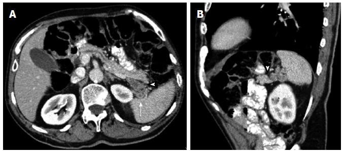
Figure 2 Axial contrast enhanced multidetector computed tomography image.
A: Depicts a nodular peripancreatic mass localized between the pancreatic tail (arrowhead) and the splenic hilum (arrow), each well separated by fat planes; B: The sagittal reformatted contrast enhanced multidetector computed tomography image allows a better identification of the surrounding fat planes (arrow and arrowhead) enabling the exclusion of a pancreatic dependency. This mass actually turned out to be a tumoral implant of a gastric neoplasm.
- Citation: Santa LGL, Retortillo JAP, Miguel AC, Klein LM. Radiology of pancreatic neoplasms: An update. World J Gastrointest Oncol 2014; 6(9): 330-343
- URL: https://www.wjgnet.com/1948-5204/full/v6/i9/330.htm
- DOI: https://dx.doi.org/10.4251/wjgo.v6.i9.330


