Copyright
©2014 Baishideng Publishing Group Inc.
World J Gastrointest Oncol. Jul 15, 2014; 6(7): 225-243
Published online Jul 15, 2014. doi: 10.4251/wjgo.v6.i7.225
Published online Jul 15, 2014. doi: 10.4251/wjgo.v6.i7.225
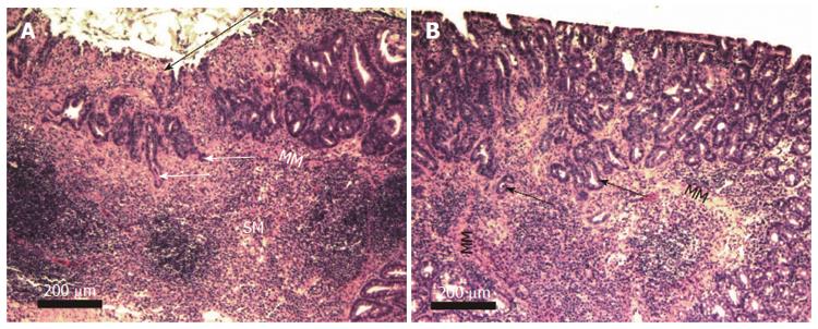
Figure 9 Two examples of mouse adenocarcinoma stage T1.
A shows a sessile serrated adenoma in the right upper portion of the image and an ulcerated region (long arrow) above an adenocarcinoma that had penetrated the muscularis mucosa. Both A and B show invasive glands (short arrows) infiltrating through the muscularis mucosa (MM) into the submucosa (SM). Images obtained with 10× objective lens.
- Citation: Prasad AR, Prasad S, Nguyen H, Facista A, Lewis C, Zaitlin B, Bernstein H, Bernstein C. Novel diet-related mouse model of colon cancer parallels human colon cancer. World J Gastrointest Oncol 2014; 6(7): 225-243
- URL: https://www.wjgnet.com/1948-5204/full/v6/i7/225.htm
- DOI: https://dx.doi.org/10.4251/wjgo.v6.i7.225


