Copyright
©2014 Baishideng Publishing Group Co.
World J Gastrointest Oncol. Mar 15, 2014; 6(3): 74-82
Published online Mar 15, 2014. doi: 10.4251/wjgo.v6.i3.74
Published online Mar 15, 2014. doi: 10.4251/wjgo.v6.i3.74
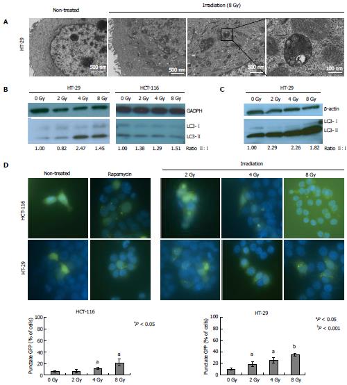
Figure 1 Radiation-induced autophagy.
A: HT-29 cells were irradiated (8 Gy) and autophagy induction was assessed by the formation of double membrane autophagosomes on transmission electron microscopy 24 h post-treatment; B: Western blot analysis of LC3 expression demonstrates increased LC3-II:I ratio and autophagy induction at 4 and 6 h post-irradiation for HT-29 and HCT116 cells, respectively; C: Increased LC3II:I ratios are seen 24 h post-irradiation at 2-8 Gy for HT-29 cells; D: GFP-labeled LC3 puncta develop within 6 h of treatment with Rapamycin (200 nmol/L) or radiation (2-8 Gy) in both HCT116 and HT-29 cell lines. Bar graphs represent quantification of percentage of cells with perinuclear punctate pattern 6 h following radiation.
- Citation: Schonewolf CA, Mehta M, Schiff D, Wu H, Haffty BG, Karantza V, Jabbour SK. Autophagy inhibition by chloroquine sensitizes HT-29 colorectal cancer cells to concurrent chemoradiation. World J Gastrointest Oncol 2014; 6(3): 74-82
- URL: https://www.wjgnet.com/1948-5204/full/v6/i3/74.htm
- DOI: https://dx.doi.org/10.4251/wjgo.v6.i3.74


