Copyright
©2014 Baishideng Publishing Group Co.
World J Gastrointest Oncol. Jan 15, 2014; 6(1): 11-21
Published online Jan 15, 2014. doi: 10.4251/wjgo.v6.i1.11
Published online Jan 15, 2014. doi: 10.4251/wjgo.v6.i1.11
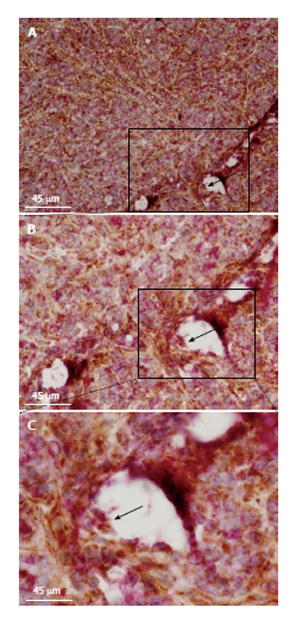
Figure 5 Tumor metabolism and malignant angiogenesis.
Histopathological images show double staining between cytochrome C oxidase (COX) and anti-CD31 antibody (clone 1A10 at 1:100; Novocastra, United States). Microvessel walls are traced with sectioned white lines (horizontal view of sectioned tumor microvessels). Black arrow indicates a microvessel lumen with double-stained cells (boxed region; transversal view of a tumor microvessel). Picture was taken at × 100 magnification and 45 μm scale bars are inserted in all images. Inset (C) shows the same boxed region at × 200 magnification. Double-stained cells are pointed out by a black arrow at the microvessel wall (inset; B). Sectioned green line circulates a niche of double-stained endothelial cells at the edge of a microvessel bifurcation. To build these images, 5 wk (20 ± 2 g) nonobese diabetic, severe combined immunodeficient mice (NOD/SCID) were subcutaneously transplanted with HT29 cells (1.5 × 10⁶ cells per mice) in agreement with the protocol approved by the Internal Animal Care, Ethical and Use Committee (n° 121/2012). All mice were acclimated for 1 wk before starting the experiment and maintained under specific pathogen-free conditions. Tumor volume was monitored throughout the whole experimental period by measures with a caliper. Mice were sacrificed under general anesthesia (1.5% Forane in 98.5% oxygen; 2l min). Tissue samples were frozen within TissueTek (Sakura, Germany) and kept at -80 °C for immunohistochemical analyses. Double-staining was performed according to our standard methods.
- Citation: Stopper H, Garcia SB, Waaga-Gasser AM, Kannen V. Antidepressant fluoxetine and its potential against colon tumors. World J Gastrointest Oncol 2014; 6(1): 11-21
- URL: https://www.wjgnet.com/1948-5204/full/v6/i1/11.htm
- DOI: https://dx.doi.org/10.4251/wjgo.v6.i1.11


